
Fatty liver disease does not arise randomly — it develops in the context of specific metabolic, dietary, genetic, and lifestyle factors that interact to produce excessive fat accumulation in the liver. Understanding what causes fatty liver disease, and what determines who progresses from simple steatosis to more severe disease, is essential for both preventing the condition from developing and for slowing or reversing it once it is established. The most common form — metabolic dysfunction-associated steatotic liver disease (MASLD) — shares its causal roots with type 2 diabetes, cardiovascular disease, and metabolic syndrome. It is fundamentally a disease of impaired metabolic homeostasis, driven by insulin resistance and its downstream consequences in hepatic lipid metabolism. This article systematically reviews the primary causes and risk factors for MASLD, explains the mechanisms through which they contribute to liver fat accumulation, and identifies which factors are most strongly associated with disease progression.
The Primary Cause: Insulin Resistance and Metabolic Dysfunction
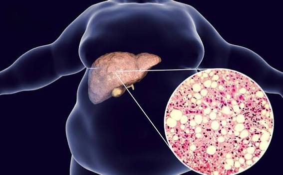
The fundamental driver of MASLD at the cellular and molecular level is insulin resistance — a state in which the body’s cells, including hepatocytes (liver cells), respond less effectively to insulin than they should. In a metabolically healthy state, insulin acts on hepatocytes to suppress de novo lipogenesis (the synthesis of new fat from glucose), promote fatty acid oxidation (the burning of fat for energy), and regulate the export of fat as VLDL particles. When hepatocytes become insulin-resistant, these regulatory processes are disrupted: insulin fails to suppress de novo lipogenesis adequately, fatty acid oxidation is impaired, and the liver receives an increased flux of free fatty acids from insulin-resistant adipose tissue (which releases more free fatty acids into the circulation when insulin signalling is impaired). The result is net fat accumulation in hepatocytes — the defining feature of MASLD. Insulin resistance is not a binary state — it exists on a spectrum — and the degree of insulin resistance correlates with the severity of hepatic steatosis. The causes of insulin resistance are multiple and interacting: obesity (particularly visceral adiposity), physical inactivity, dietary pattern (high refined carbohydrate and saturated fat intake), genetics, ageing, sleep disruption, and psychological stress all contribute. Understanding insulin resistance as the central mechanism of MASLD helps make sense of why the condition shares its risk factors so completely with type 2 diabetes and cardiovascular disease: they are all metabolic consequences of the same underlying pathophysiology. The MASLD diagnostic criteria formalise this connection by requiring at least one cardiometabolic risk factor alongside hepatic steatosis for the diagnosis to be made.
Obesity and Body Composition
Obesity is the single most important and most prevalent risk factor for MASLD. The prevalence of MASLD in people with obesity (BMI above 30 kg/m²) is approximately sixty to eighty percent — compared with fifteen to twenty percent in the general adult population. However, the relationship between obesity and MASLD is more nuanced than total body weight alone suggests. Visceral adiposity — fat stored within the abdominal cavity, around the intra-abdominal organs — is more strongly associated with MASLD than total body fat, even after adjusting for BMI. Visceral adipose tissue is more metabolically active than subcutaneous fat and releases more free fatty acids into the portal circulation, delivering a higher lipid load directly to the liver. Visceral adiposity is measured clinically by waist circumference (above 102 cm in men and 88 cm in women in European populations; lower thresholds apply in Asian populations) or by imaging. Lean MASLD: Approximately fifteen to twenty percent of MASLD cases occur in patients with a normal BMI (below 25 kg/m²) — a phenotype known as lean MASLD. These patients often have elevated visceral adiposity relative to their body weight (a high waist-to-hip ratio, suggesting preferential visceral fat distribution despite normal BMI), and carry the same cardiometabolic risk factors as obese MASLD patients, though the metabolic burden may be less severe. Lean MASLD is more common in Asian populations, where MASLD develops at lower BMI thresholds than in European populations due to differences in body fat distribution. The relationship between weight loss and MASLD: Intentional weight loss of five percent of body weight is associated with significant reduction in hepatic steatosis; ten percent weight loss with MASH resolution; fifteen to twenty percent weight loss with fibrosis regression. This dose-response relationship between weight loss and liver improvement is one of the most consistently replicated findings in MASLD research and forms the basis for lifestyle intervention recommendations in all major guidelines.
Type 2 Diabetes and Insulin Resistance Syndromes
Type 2 diabetes is present in approximately fifty to seventy percent of patients with MASLD — and conversely, MASLD is present in approximately fifty to seventy percent of patients with type 2 diabetes. The relationship is bidirectional and mechanistically coupled: the insulin resistance that causes type 2 diabetes also drives hepatic steatosis, and MASLD in turn worsens hepatic insulin resistance and contributes to hyperglycaemia. MASLD with type 2 diabetes carries a substantially higher risk of progression to MASH and advanced fibrosis than MASLD without diabetes — making diabetes one of the most important modifiable risk factors for severe liver disease. The glycaemic treatment of type 2 diabetes influences MASLD trajectory: pioglitazone (a thiazolidinedione) has the strongest evidence for histological improvement in MASLD among glucose-lowering agents; GLP-1 receptor agonists (semaglutide, liraglutide) demonstrate significant liver steatosis and fibrosis reduction in clinical trials; SGLT2 inhibitors show consistent reductions in liver fat and enzymes. Metformin does not produce consistent histological improvement in MASLD despite its role in glycaemic management. Pre-diabetes and insulin resistance without overt diabetes are also significant MASLD risk factors. Impaired fasting glucose or impaired glucose tolerance increases MASLD risk significantly, and patients with metabolic syndrome (three or more of: central obesity, hypertension, hyperglycaemia, elevated triglycerides, low HDL cholesterol) have MASLD prevalence rates of over fifty percent in population studies. The diagnosis of metabolic syndrome is therefore a high-yield trigger for proactive fatty liver assessment.
Dyslipidaemia and Lipid Metabolism Abnormalities
Lipid abnormalities are both a cause and a consequence of MASLD. The characteristic dyslipidaemia pattern associated with MASLD — elevated triglycerides and low HDL cholesterol — reflects the same insulin resistance state that drives hepatic steatosis. Elevated triglycerides (above 1.7 mmol/L or 150 mg/dL) indicate impaired clearance of VLDL particles from the circulation and are a consistent predictor of MASLD prevalence and severity. Low HDL (below 1.0 mmol/L in men or 1.3 mmol/L in women) reflects impaired reverse cholesterol transport. LDL cholesterol is less consistently elevated in MASLD than triglycerides and HDL, and the LDL particles in MASLD patients are often small and dense (more atherogenic). The liver’s impaired ability to process lipids correctly — reflected in the dyslipidaemia — is both a marker of the underlying insulin resistance and an independent contributor to hepatic steatosis through increased intrahepatic lipid availability. Treating dyslipidaemia — particularly elevated triglycerides — with lifestyle modification (dietary carbohydrate reduction, physical activity, omega-3 fatty acids) contributes to hepatic steatosis reduction. Statins, despite historical concerns about their use in liver disease, are safe and beneficial in MASLD patients and are now recommended for cardiovascular risk management in MASLD — they reduce liver enzyme levels and may have anti-fibrotic effects.
Dietary Factors
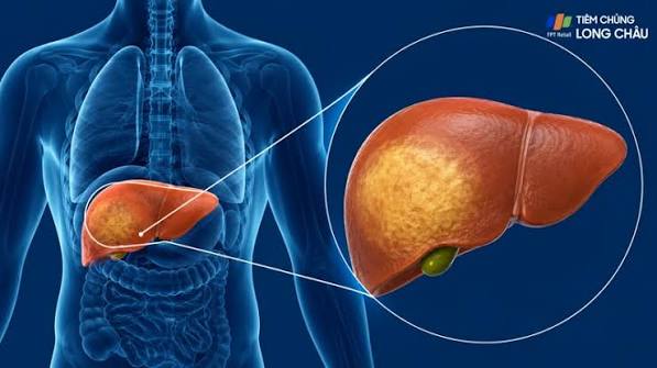
Diet is one of the most modifiable contributors to MASLD risk. The key dietary factors associated with MASLD development and severity include: Excess caloric intake and weight gain — the most impactful dietary variable, as hepatic steatosis tracks closely with energy surplus and weight. High fructose intake: Fructose, unlike glucose, is almost exclusively metabolised by the liver and is a potent inducer of de novo lipogenesis. High fructose consumption — particularly from sugar-sweetened beverages (SSBs) — is associated with MASLD prevalence and steatosis severity independent of total calorie intake. A meta-analysis of prospective cohort studies found that SSB consumption was associated with a significantly elevated MASLD risk, and the association was stronger for fructose-containing SSBs than for diet alternatives. Saturated fat: High saturated fat intake promotes hepatic steatosis and MASH activity through multiple mechanisms including ceramide synthesis, endoplasmic reticulum stress, and TLR4 activation. Refined carbohydrates: Rapidly absorbed carbohydrates (white bread, white rice, processed cereals) stimulate insulin secretion and de novo lipogenesis more than equivalent calories from whole grains or vegetables, contributing to insulin resistance and hepatic fat accumulation. Ultra-processed foods (UPF): Emerging evidence links high UPF consumption with MASLD prevalence and severity, though this may in part reflect the high content of refined carbohydrates, saturated fat, and fructose in these foods. Dietary patterns associated with reduced MASLD risk: The Mediterranean diet — high in olive oil, vegetables, whole grains, legumes, and fish; low in red and processed meat and refined carbohydrates — consistently demonstrates hepatic steatosis reduction in randomised controlled trials, independently of caloric restriction. Coffee consumption (three or more cups per day) is associated with reduced liver fibrosis in MASLD, attributed to the anti-inflammatory and antioxidant properties of polyphenols and caffeine.
Physical Inactivity
Sedentary behaviour and low cardiorespiratory fitness are independent risk factors for MASLD, even after adjusting for BMI and metabolic risk factors. Skeletal muscle is the primary site of insulin-stimulated glucose uptake — reduced muscle mass and impaired muscle metabolism in physically inactive individuals contributes to insulin resistance and increased hepatic lipid delivery. Regular aerobic exercise reduces hepatic steatosis through several mechanisms: direct stimulation of hepatic and skeletal muscle fatty acid oxidation, improvement of whole-body insulin sensitivity, reduction in visceral adiposity, and reduction in de novo lipogenesis. Studies show that moderate aerobic exercise (one hundred and fifty minutes per week) produces significant CAP score reduction on FibroScan and ALT reduction in MASLD patients, independent of weight loss. Resistance training (strength exercise) also reduces hepatic steatosis and improves metabolic syndrome parameters, and may be particularly valuable for patients who cannot tolerate sustained aerobic exercise. The combination of aerobic and resistance training is more effective than either alone for reducing liver fat.
Genetic Risk Factors
Genetic variation plays a significant role in determining individual susceptibility to MASLD and the severity of disease for a given metabolic burden. The most extensively characterised genetic risk variant is the PNPLA3 I148M polymorphism: carriers of the M148 allele (approximately a quarter of European and half of Hispanic populations) have higher hepatic fat content, higher risk of MASH and fibrosis, and higher risk of HCC than non-carriers with equivalent metabolic risk profiles. The PNPLA3 protein (patatin-like phospholipase domain containing protein 3) is involved in hepatic triglyceride hydrolysis and lipid droplet remodelling, and the I148M variant reduces its lipase activity, impairing hepatic lipid clearance. The TM6SF2 E167K variant is associated with higher hepatic fat content and liver enzyme elevation but, paradoxically, a lower risk of cardiovascular disease — because TM6SF2 is involved in VLDL secretion, and the variant reduces VLDL export from the liver (retaining lipids intrahepactically) while also reducing circulating atherogenic lipoproteins. The MBOAT7 rs641738 variant is associated with increased MASLD susceptibility and fibrosis risk, particularly in patients of European ancestry. Genetic testing for MASLD susceptibility variants is not currently part of routine clinical practice but is an active area of research — the development of a validated polygenic risk score for MASLD progression could eventually inform personalised surveillance intensity and pharmacological treatment thresholds. Family history of liver disease, particularly in first-degree relatives, should be elicited and considered when assessing MASLD risk.
Alcohol and Other Secondary Causes
While MASLD is defined as occurring in the absence of clinically significant alcohol use, alcohol is worth addressing because of its frequent coexistence with metabolic risk factors. Any alcohol consumption adds a superimposed hepatic injury to the existing metabolic liver disease in MASLD patients — the MetALD category in the 2023 terminology update recognises patients with both metabolic dysfunction and significant alcohol use as a distinct high-risk group. Secondary causes of fatty liver disease that should be excluded during the diagnostic workup include: drug-induced liver disease (corticosteroids, amiodarone, tamoxifen, methotrexate, valproate, and other medications cause hepatic steatosis); hypothyroidism (impairs hepatic fatty acid oxidation); coeliac disease (associated with liver enzyme elevation); Wilson’s disease (in younger patients); and total parenteral nutrition. These secondary causes are excluded as part of the standard MASLD diagnostic pathway and their identification changes the management approach entirely.
Frequently Asked Questions About Fatty Liver Causes
Can you get fatty liver disease without being overweight?
Yes — approximately fifteen to twenty percent of MASLD cases occur in patients with a normal BMI, a phenotype known as lean MASLD. These patients typically have elevated visceral adiposity relative to their total body weight, insulin resistance, and other metabolic risk factors despite a normal BMI. Lean MASLD is more common in Asian populations, where fatty liver develops at lower BMI thresholds. The absence of obesity does not exclude MASLD — liver blood test assessment is warranted in any patient with metabolic risk factors or unexplained liver enzyme elevation, regardless of weight.
Is fatty liver disease hereditary?
There is a significant genetic component to MASLD susceptibility and severity — the PNPLA3 I148M variant, TM6SF2 E167K variant, and MBOAT7 variant all influence hepatic fat accumulation and fibrosis risk. However, genetic variants do not cause MASLD in isolation — they modify the metabolic and dietary context within which MASLD develops. A carrier of the high-risk PNPLA3 genotype who maintains a healthy weight, exercises regularly, and has well-controlled metabolic risk factors has a substantially lower MASLD risk than a carrier who is obese and insulin-resistant. The genetic risk increases susceptibility and lowers the metabolic threshold at which MASLD develops, but lifestyle management remains the primary determinant of disease development and progression for most patients.
Does drinking too much sugar cause fatty liver?
High fructose consumption — particularly from sugar-sweetened beverages — is a significant and well-documented contributor to MASLD, through its role as a substrate for hepatic de novo lipogenesis. Fructose is almost exclusively metabolised by the liver and, at high consumption levels, delivers a lipogenic substrate load that exceeds the liver’s capacity to oxidise or export it, leading to fat accumulation. However, total calorie excess and insulin resistance from any dietary source are more powerful determinants of MASLD than fructose specifically. Reducing sugar-sweetened beverage consumption is one of the most high-impact dietary changes available to patients with MASLD, both for its direct hepatic effect and for its contribution to overall caloric balance and insulin sensitivity. The FibroScan guide and dedicated fatty liver dietary articles in this series provide practical dietary guidance.
Sources: EASL–EASD–EASO — MASLD Clinical Practice Guidelines · AASLD — Liver Disease Clinical Guidance · NIDDK — NAFLD and NASH
Hypertension and Cardiovascular Risk Factors
Hypertension is an independent risk factor for MASLD and for fibrosis progression within MASLD — present in approximately sixty to seventy percent of patients with advanced MASLD-related fibrosis in clinical series. The mechanistic links between hypertension and MASLD are multiple: shared insulin resistance as a common upstream driver; activation of the renin-angiotensin system (RAS), which promotes hepatic stellate cell activation and fibrogenesis directly; endothelial dysfunction affecting the hepatic microvasculature; and oxidative stress in both the vascular and hepatic compartments. Angiotensin II, the main effector of the RAS, is a potent pro-fibrotic mediator in the liver — a finding that has driven interest in RAS inhibitors (ACE inhibitors and ARBs) as potential anti-fibrotic agents in MASLD, though clinical trial evidence for histological benefit remains limited. The coexistence of MASLD and hypertension substantially elevates cardiovascular risk above either condition alone — patients with both conditions should be under active cardiovascular risk management in parallel with hepatic monitoring. The aggregate cardiovascular risk in a patient with MASLD, type 2 diabetes, hypertension, and dyslipidaemia — all components of metabolic syndrome — is markedly elevated, and cardiovascular disease remains the leading cause of death in MASLD cohort studies, ahead of liver-related complications at the population level.
Sleep Disorders and Circadian Disruption
Sleep disorders — particularly obstructive sleep apnoea (OSA) — are emerging as significant independent risk factors for MASLD severity and fibrosis progression. OSA is highly prevalent in patients with obesity and MASLD (estimated forty to eighty percent prevalence in MASLD clinic populations), and the relationship is bidirectional: obesity promotes OSA through mechanical effects on the upper airway, and OSA worsens insulin resistance, promotes weight gain, and may directly injure the liver through intermittent hypoxaemia. Intermittent hypoxia — the recurrent oxygen desaturation events characteristic of OSA — activates hypoxia-inducible factor 1-alpha (HIF-1α) in hepatocytes, promoting de novo lipogenesis and suppressing fatty acid oxidation, and activates hepatic stellate cells through NF-κB and TGF-β pathways, promoting fibrogenesis. Several prospective studies have found that OSA severity (as measured by the apnoea-hypopnoea index) correlates with hepatic steatosis grade and fibrosis stage in MASLD patients, independent of BMI and metabolic risk factors. CPAP (continuous positive airway pressure) treatment for OSA has been shown to reduce liver enzyme levels and improve insulin sensitivity in obese patients, though evidence for histological improvement in MASLD is preliminary. Shift work and sleep restriction are also associated with increased MASLD risk through circadian disruption of hepatic metabolic rhythms: the liver expresses robust circadian rhythms in lipid and glucose metabolism, and chronic circadian disruption (as in shift workers) impairs these rhythms and promotes hepatic steatosis. Patients with MASLD and suspected sleep disorders should be assessed for OSA, as treatment of OSA is a clinically meaningful intervention with metabolic and potentially hepatic benefits beyond its primary indication.
The Ageing Liver and Increased Risk With Age
Age is an independent risk factor for MASLD — both for its development and for fibrosis progression once established. MASLD prevalence increases progressively with age, from approximately fifteen percent in young adults to over thirty percent in those aged sixty and above. Several age-related changes contribute: mitochondrial function declines with age, impairing hepatic fatty acid oxidation; insulin sensitivity decreases with age independent of weight gain; the composition of gut microbiota changes with age; and accumulated exposure to metabolic risk factors over decades increases the probability of hepatic steatosis and fibrosis. Older patients with MASLD are at higher risk of advanced fibrosis than younger patients with equivalent metabolic profiles, and age above fifty is incorporated into the FIB-4 index as an independent predictor of fibrosis probability — the formula uses age as a multiplier, meaning that the same ALT, AST, and platelet values produce a higher FIB-4 score in an older patient. In post-menopausal women, the loss of oestrogen’s hepatoprotective effects contributes to the convergence of MASLD risk between men and women — pre-menopausal women have lower MASLD prevalence than age-matched men, but post-menopausal women approach male MASLD prevalence rates. The implication for clinical practice is that age itself is a reason to assess liver health proactively: a sixty-year-old patient with metabolic syndrome has a higher pre-test probability of advanced fibrosis than a forty-year-old with the same metabolic profile, and the investigation threshold should be calibrated accordingly. Identifying and managing MASLD in older adults before cirrhosis is established is particularly important because the regenerative capacity of the ageing liver is reduced, and recovery from advanced liver disease is more limited than in younger patients.
Understanding your personal risk factor profile is the first step in engaging with the liver function test monitoring and non-invasive fibrosis assessment pathway. For patients with type 2 diabetes or metabolic syndrome, proactive engagement with abdominal ultrasound and FIB-4 calculation — as recommended by the EASL and AASLD guidelines — identifies the subset of patients with occult advanced fibrosis who require specialist hepatology management and more intensive metabolic risk factor intervention. The practical implication is that every modifiable risk factor addressed — through weight management, glycaemic control, lipid lowering, blood pressure treatment, alcohol reduction, increased physical activity — simultaneously reduces the MASLD progression risk and the cardiometabolic risk that coexists with it.
I always assumed fatty liver was just about eating too much fat. This article changed my understanding completely — the insulin resistance explanation makes much more sense, especially since I have type 2 diabetes. The section on fructose and sugar-sweetened drinks was particularly eye-opening.
Thank you Marcus — the insulin resistance framing is really the key to understanding why MASLD is so closely linked to type 2 diabetes, and why improving glycaemic control benefits both conditions simultaneously. The fructose piece is one of the most actionable dietary changes available: reducing or eliminating sugar-sweetened beverages is often the single highest-impact dietary intervention for someone managing both T2D and MASLD, because it addresses both fructose load and overall caloric intake in one step. The dedicated fatty liver diet article in this series covers the practical dietary changes in more detail.
The sleep apnoea connection surprised me. I’ve had OSA for years and was recently diagnosed with MASLD — I didn’t realise they could be linked. I’ll mention this to my hepatologist at my next appointment. Good article, very informative.