
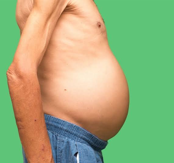
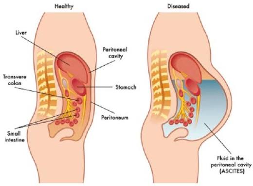
A swollen belly — abdominal distension or bloating — has many causes ranging from the entirely benign (gas, constipation, overeating) to the medically serious (ascites, organomegaly, or abdominal masses). Among the serious causes, liver disease occupies a central position: ascites — the accumulation of fluid in the peritoneal cavity — is the most common complication of cirrhosis and one of the most recognizable signs of advanced liver disease. A patient who develops a progressively swollen belly over weeks to months alongside profound fatigue, jaundice, or a history of liver disease is very likely experiencing the onset of ascites — a development that transforms the natural history of cirrhosis from compensated (asymptomatic) to decompensated and substantially worsens long-term prognosis.
Ascites is not merely an inconvenience. It signals that the liver has lost sufficient function and architecture to maintain normal fluid balance, that portal hypertension has developed to a degree sufficient to drive fluid transudation into the peritoneum, and that the kidneys and neurohumoral systems regulating fluid retention are responding to perceived hypovolemia with avid sodium and water retention. The median survival after first presentation of ascites in cirrhosis is approximately two years, substantially worse than for patients with compensated cirrhosis who have no clinical decompensation. Understanding ascites — its mechanism, its complications, its treatment options, and the warning signs of deterioration — is essential for both patients with liver disease and the clinicians who care for them.
How Cirrhosis Causes Ascites — Portal Hypertension and Sodium Retention
The pathophysiology of cirrhotic ascites integrates portal hemodynamics, hepatic function, renal physiology, and neurohumoral regulation into a self-reinforcing cycle of fluid retention. The primary driver is portal hypertension — elevated pressure in the portal venous system from the increased resistance to blood flow through the fibrosed cirrhotic liver. Portal hypertension causes splanchnic vasodilation (dilation of blood vessels in the intestinal circulation) mediated by nitric oxide and other vasodilatory mediators produced in response to high portal pressure. Splanchnic vasodilation reduces effective circulatory volume — even though total blood volume may be normal or increased, the blood is pooled in the dilated splanchnic circulation rather than effectively perfusing vital organs.
The kidney interprets the reduced effective circulatory volume as hypovolemia and activates the renin-angiotensin-aldosterone system (RAAS), antidiuretic hormone (ADH), and sympathetic nervous system — all of which promote avid sodium and water retention. The retained sodium and water cannot remain in the intravascular compartment because of the reduced oncotic pressure from low serum albumin (produced by the failing liver) and the elevated hydrostatic pressure in the portal system; instead, fluid transudates across the peritoneal surface and accumulates as ascites. The result is a vicious cycle in which the liver’s failure to clear vasoactive substances maintains splanchnic vasodilation, the kidneys continue to retain sodium and water in a futile attempt to correct apparent hypovolemia, and the peritoneal cavity fills progressively with fluid.
The serum albumin-ascites gradient (SAAG) — calculated as serum albumin minus ascites albumin — is the most reliable diagnostic test for distinguishing ascites caused by portal hypertension (SAAG ≥ 1.1 g/dL, indicating high protein transudation) from ascites caused by malignancy, peritoneal tuberculosis, or other non-portal-hypertension mechanisms (SAAG < 1.1 g/dL). All patients with new-onset ascites should undergo diagnostic paracentesis — a needle aspiration of ascitic fluid — to confirm the SAAG and to exclude spontaneous bacterial peritonitis (SBP), a potentially life-threatening infection of the ascitic fluid that can occur without preceding fever or abdominal pain and requires prompt diagnosis and treatment.
Clinical Features of Ascites — Symptoms, Signs, and Grading
Small-volume ascites (Grade 1) — detectable only on ultrasound, not clinically apparent — produces no symptoms and may not require specific treatment beyond sodium restriction and management of the underlying liver disease. Moderate ascites (Grade 2) produces visible abdominal distension, a sensation of abdominal fullness or heaviness, and shifting dullness on percussion (dullness that shifts with patient positioning as the fluid redistributes under gravity). Large or tense ascites (Grade 3) causes significant abdominal discomfort, early satiety from gastric compression, breathlessness from elevation of the diaphragm, difficulty walking from the weight of the distended abdomen, and an everted umbilicus from the outward pressure of the fluid. In the most severe cases, the abdomen is taut and tender, and the overlying skin may be stretched to translucency.
Peripheral edema — swelling of the ankles and legs — frequently accompanies ascites in decompensated cirrhosis, reflecting the same underlying mechanisms of reduced oncotic pressure and sodium retention. Hepatic hydrothorax — the passage of ascitic fluid through small diaphragmatic defects into the pleural cavity — affects approximately five percent of patients with cirrhotic ascites and produces right-sided pleural effusion that can cause significant breathlessness disproportionate to the degree of abdominal fluid. Umbilical hernias develop in a substantial minority of patients with chronic ascites from the persistent outward pressure; they are prone to incarceration and rupture in patients with tense ascites and require careful surgical planning in the context of liver disease-related coagulopathy and surgical risk. The presence of caput medusae — dilated veins radiating from the umbilicus over the abdominal wall — indicates significant portal hypertension and is a distinctive physical sign of cirrhotic decompensation.
Treatment of Cirrhotic Ascites — Sodium Restriction, Diuretics, and Paracentesis
The cornerstone of ascites management is dietary sodium restriction — limiting intake to less than 2000 milligrams of sodium per day (approximately 5 grams of table salt) — combined with diuretic therapy to achieve a negative sodium balance and fluid loss. Spironolactone — an aldosterone antagonist that blocks the aldosterone-driven sodium retention that drives ascites — is the first-line diuretic for cirrhotic ascites, typically initiated at 100 milligrams daily and titrated to 400 milligrams daily as needed. Furosemide is added at a starting dose of 40 milligrams daily (with a spironolactone:furosemide ratio of 100:40 maintained through titration) to augment natriuresis. The target weight loss during diuresis is approximately 500 grams per day in patients without peripheral edema and 1000 grams per day in those with both ascites and edema — more rapid fluid removal risks precipitating renal impairment and electrolyte disturbances.
Large-volume paracentesis (LVP) — needle drainage of ascitic fluid — is indicated for tense or refractory ascites that does not respond to maximum-dose diuretics, or as a rapid intervention for symptomatic relief in patients with respiratory compromise or severe abdominal discomfort. Removal of more than five liters of ascitic fluid in a single session is associated with post-paracentesis circulatory dysfunction (PPCD), a hemodynamic instability that activates the RAAS and worsens long-term prognosis; intravenous albumin infusion at a dose of 6–8 grams per liter of ascites removed (above five liters) is standard practice to prevent PPCD. Patients requiring LVP more than twice per month despite maximum medical therapy have refractory ascites, which carries a particularly poor prognosis — median survival under twelve months — and should be evaluated for liver transplantation and for transjugular intrahepatic portosystemic shunt (TIPS) placement if transplantation is not immediately available.
Spontaneous Bacterial Peritonitis — A Life-Threatening Complication
Spontaneous bacterial peritonitis (SBP) occurs when bacteria colonize the ascitic fluid — most commonly Escherichia coli, Klebsiella pneumoniae, or Streptococcus pneumoniae — without an identifiable intra-abdominal source of infection. It is a serious complication with an in-hospital mortality of fifteen to twenty percent even with appropriate antibiotic treatment, and survivors have a one-year mortality rate exceeding fifty percent from renal failure and progressive liver disease. SBP may present without fever or abdominal pain in a significant proportion of patients — the first manifestation is sometimes a change in mental status (hepatic encephalopathy), unexplained worsening of renal function, or simply a clinical deterioration in a patient with known ascites. Diagnostic paracentesis with an ascitic fluid polymorphonuclear neutrophil (PMN) count above 250 cells per cubic millimeter confirms the diagnosis, and antibiotic treatment — typically intravenous cefotaxime or a third-generation cephalosporin — should be initiated empirically while culture results are pending. Intravenous albumin at diagnosis reduces the risk of hepatorenal syndrome and improves survival. Secondary prophylaxis with norfloxacin or trimethoprim-sulfamethoxazole is recommended after the first SBP episode to prevent recurrence.
Non-Cirrhotic Causes of Abdominal Swelling to Exclude
Not every swollen belly in a patient with liver disease is ascites, and not every ascites is from cirrhosis. Malignant ascites — from peritoneal metastases of colorectal, ovarian, gastric, or pancreatic cancer — produces a SAAG below 1.1 g/dL, indicating that the fluid accumulation results from peritoneal tumor involvement rather than portal hypertension. Malignant ascites often accumulates rapidly, may be blood-tinged, and carries a grim prognosis; peritoneal cytology and imaging are required to establish the diagnosis. Tuberculous peritonitis produces ascites in patients with disseminated tuberculosis or with reactivation of latent TB in immunosuppressed or malnourished individuals; the SAAG is low, and ascitic fluid analysis showing lymphocyte predominance and elevated adenosine deaminase (ADA) supports the diagnosis.
Cardiac ascites — from right heart failure, constrictive pericarditis, or Budd-Chiari syndrome (hepatic vein thrombosis) — produces a high SAAG (≥ 1.1 g/dL, mimicking cirrhotic ascites) and requires imaging of the hepatic veins and cardiac evaluation to distinguish from cirrhosis. Budd-Chiari syndrome is particularly important to consider in young patients with unexplained ascites and hepatomegaly, especially in the context of myeloproliferative disorders, thrombophilias, or oral contraceptive use — conditions that increase thrombotic risk. The distinction has direct treatment implications: anticoagulation and, in some cases, TIPS or liver transplantation are the treatments for Budd-Chiari, rather than the diuretic regimens used for cirrhotic portal hypertension. Pancreatic ascites from ductal disruption or pseudocyst rupture — elevated ascitic fluid amylase is the diagnostic finding — and chylous ascites from lymphatic disruption are other non-cirrhotic causes of abdominal swelling that require specific investigation and management strategies distinct from portal-hypertensive ascites.
Frequently Asked Questions About Swollen Belly and Liver Disease
Is all abdominal swelling in liver disease from ascites?
No. Hepatomegaly (an enlarged liver) and splenomegaly (an enlarged spleen from portal hypertension) can both cause abdominal fullness and visible abdominal distension, particularly in the upper abdomen. Constipation — which is common in patients with liver disease taking lactulose or with reduced physical activity — contributes to abdominal discomfort and bloating. Gas from altered gut motility in cirrhosis can cause significant abdominal bloating that is not fluid. Clinical examination, particularly percussion for shifting dullness and fluid wave, distinguishes ascites (which shifts) from gas, stool, or solid organomegaly (which do not shift). Ultrasound confirms the presence of free fluid and quantifies its volume more precisely than clinical examination.
Can ascites be reversed?
Ascites from cirrhosis is reversible in some patients when the underlying liver disease is successfully treated. In viral hepatitis-related cirrhosis, achieving sustained virological response (SVR) with direct-acting antivirals for HCV or effective suppression of HBV can lead to meaningful improvement in liver function and, in some patients, resolution of ascites as portal pressure declines with fibrosis regression. In alcoholic cirrhosis, sustained abstinence from alcohol allows hepatic inflammation to resolve and, in patients whose liver disease has not progressed to an irreversible stage, may produce progressive improvement in liver function including resolution of ascites over months to years. Patients with refractory ascites and end-stage liver disease, however, will not achieve reversal without liver transplantation. The development of ascites is a key inflection point in the decision to refer a patient for transplantation evaluation.
What are the signs that ascites is becoming dangerous?
Warning signs requiring prompt evaluation include: fever with abdominal pain or tenderness (possible SBP); confusion or altered mental status in a patient with known ascites (possible SBP triggering hepatic encephalopathy, or HE from another cause); rapid increase in abdominal girth suggesting rapid fluid accumulation; breathlessness from elevation of the diaphragm or hepatic hydrothorax; leg swelling that is worsening or has become asymmetric (possible deep vein thrombosis); and development of dark urine or worsening jaundice alongside the ascites. Any of these features should prompt immediate contact with the treating hepatologist or, if unavailable, emergency department evaluation.
Sources: NIDDK — Cirrhosis · ACG — Ascites · Mayo Clinic — Ascites
Hepatorenal Syndrome — When Ascites Leads to Kidney Failure
Hepatorenal syndrome (HRS) is one of the most feared complications of cirrhotic ascites — a functional kidney failure that develops in response to the hemodynamic derangements of advanced liver disease without any intrinsic structural renal pathology. In HRS, the kidneys are structurally intact but are not perfused adequately because of the profound splanchnic vasodilation and compensatory renal vasoconstriction that characterize advanced portal hypertension. HRS type 1 (now classified as AKI-HRS under modern criteria) is a rapidly progressive form that develops over less than two weeks, often precipitated by SBP, gastrointestinal bleeding, or overly aggressive diuresis; it carries a mortality rate exceeding fifty percent without treatment. HRS type 2 (now classified as CKD-HRS) is a slower, more chronic kidney impairment associated with refractory ascites and a worse long-term prognosis than ascites without kidney involvement.
Treatment of HRS prioritizes restoring effective circulatory volume and reversing renal vasoconstriction. The combination of terlipressin (a vasopressin analogue that reduces splanchnic vasodilation) with albumin infusion has demonstrated improved renal function and survival in AKI-HRS in clinical trials and is the standard of care in countries where terlipressin is available. Norepinephrine infusion combined with albumin is an alternative where terlipressin is not available. Midodrine (an oral alpha-1 adrenergic agonist) combined with octreotide (a somatostatin analogue) and albumin is a less potent but more widely available regimen used in some centers. Renal replacement therapy (dialysis) may bridge patients to liver transplantation but does not reverse the underlying mechanism. Liver transplantation — ideally combined liver-kidney transplantation in patients with established CKD-HRS — remains the definitive treatment and provides the best long-term outcomes.
TIPS — Transjugular Intrahepatic Portosystemic Shunt for Refractory Ascites
TIPS — a percutaneous procedure in which a shunt is created between the portal vein and a hepatic vein through the liver parenchyma using a covered metal stent — reduces portal pressure by creating a direct bypass of the high-resistance cirrhotic liver. By lowering portal hypertension, TIPS reduces the splanchnic vasodilation that drives ascites, allowing the kidney to reduce its avid sodium retention and enabling the ascites to resolve or be better controlled with diuretics. TIPS is effective at controlling refractory ascites in the majority of patients who receive it — approximately seventy to eighty percent achieve ascites resolution or a significant reduction in the need for paracentesis — and may improve survival compared to repeated large-volume paracentesis in selected patients.
However, TIPS carries significant risks that must be weighed against its benefits in individual patients. By diverting portal blood flow away from the liver and into the systemic circulation, TIPS increases the ammonia and other nitrogen-containing substances reaching the brain, worsening or precipitating overt hepatic encephalopathy (HE) in approximately thirty percent of patients. Covert HE (mild cognitive impairment without overt confusion) is even more common after TIPS. Careful patient selection — excluding those with pre-existing severe encephalopathy, bilirubin above three milligrams per deciliter, or advanced hepatic reserve impairment (Child-Pugh C with score above twelve) — reduces but does not eliminate this risk. Prophylactic lactulose or rifaximin is often prescribed after TIPS to reduce ammonia production and minimize the risk of post-TIPS encephalopathy. TIPS does not replace liver transplantation for patients with suitable indications — it is a bridge procedure intended to reduce complications and maintain quality of life while awaiting a suitable organ.
Liver Transplantation — The Definitive Treatment for Decompensated Cirrhosis
Liver transplantation is the definitive treatment for decompensated cirrhosis with ascites and the only intervention that addresses the underlying cause rather than managing the consequences. The development of ascites — particularly refractory ascites requiring repeated paracentesis — is one of the key clinical events that triggers transplantation evaluation, and MELD (Model for End-Stage Liver Disease) score — calculated from creatinine, bilirubin, and INR — determines organ allocation priority in most countries. The MELD-Na score, which incorporates serum sodium (a marker of dilutional hyponatremia and advanced circulatory dysfunction), has replaced the original MELD score in the United States and several other countries because it more accurately predicts ninety-day waitlist mortality in patients with ascites and hyponatremia.
Patients with ascites who are being evaluated for liver transplantation should be maintained in the best possible clinical condition — optimizing nutrition (which is frequently impaired in cirrhosis from anorexia, malabsorption, and reduced hepatic protein synthesis), treating infections promptly, maintaining diuretic therapy and sodium restriction, and monitoring for complications including SBP, HRS, and hepatic encephalopathy. Palliative care involvement is appropriate for patients who are not transplant candidates, focusing on symptom management — including serial paracentesis for refractory ascites, treatment of pruritus and encephalopathy, and careful management of the psychosocial burden of advanced liver disease. The development of ascites marks a point in the trajectory of liver disease where honest, compassionate conversations about prognosis, transplantation eligibility, and end-of-life preferences are both clinically necessary and ethically important.
The daily lived experience of patients managing cirrhotic ascites and its complications is substantially more challenging than the clinical management algorithms suggest. Fluid and sodium restriction are difficult to maintain, particularly for patients with reduced appetites, taste changes from zinc deficiency, or limited food preparation resources. Monitoring weight daily — the most practical proxy for fluid balance at home — requires reliable scales and patient education about the meaning of weight changes. A weight gain of more than two kilograms over two to three days should prompt contact with the clinical team for possible diuretic adjustment, while rapid weight loss during diuresis (more than five hundred grams per day in patients without peripheral edema) should trigger diuretic dose reduction to prevent prerenal acute kidney injury. The sodium restriction required to control ascites (less than two grams per day) is significantly tighter than general low-sodium dietary advice and may require dietitian support to implement practically, particularly in patients who rely on processed foods, eat outside the home frequently, or have cultural dietary patterns that are high in sodium. The combination of hepatic encephalopathy (which impairs cognition and judgment), fatigue, anorexia, and the social disruption of chronic illness creates a challenging environment for the self-management behaviors that prevent complications and hospitalizations. Structured patient education — ideally delivered by a hepatology nurse specialist or advanced practice provider with dedicated time for this purpose — demonstrably reduces readmission rates and improves quality of life in patients with cirrhotic decompensation, and is an underinvested component of chronic liver disease management in many healthcare systems.
For patients and families navigating a new diagnosis of cirrhotic ascites, the volume of information required for safe self-management — sodium restriction targets, daily weighing, diuretic adjustments, warning signs of SBP and HRS, and the role of transplantation evaluation — can be overwhelming. A practical first step is to request a dedicated appointment with a hepatology nurse or dietitian for tailored education, rather than relying solely on information received during an acute hospital admission. Understanding that ascites management is a dynamic process — requiring frequent dose adjustments, dietary adherence, and prompt reporting of changes — rather than a static prescription helps patients engage as active partners in their care. Connecting with peer support groups for people with cirrhosis can also reduce the isolation associated with managing a serious chronic condition, and many hepatology centers can provide referrals to these resources. The long-term outlook for patients with cirrhotic ascites has improved meaningfully with advances in transplantation medicine, antiviral therapy for hepatitis B and C, and the expanding use of TIPS — reasons for cautious optimism alongside the ongoing challenges of managing decompensated liver disease.