
Celiac disease is an autoimmune condition triggered by gluten — a protein found in wheat, barley, and rye — in which the immune system mounts an abnormal response to gluten peptides in the small intestinal mucosa, causing villous atrophy (destruction of the absorptive villi that line the small intestine), malabsorption, and systemic complications. It affects approximately one percent of the global population but remains significantly underdiagnosed — studies consistently show that for every one person diagnosed with celiac disease, six to ten have the condition but remain undiagnosed. This diagnostic gap exists because celiac disease is extraordinarily protean in its clinical presentation: the classic picture of severe diarrhoea, weight loss, and malabsorption in a symptomatic child is only one face of a disease that more often presents in adults with subtle or entirely atypical symptoms including fatigue, iron deficiency anaemia, unexplained osteoporosis, infertility, neurological symptoms, dermatitis herpetiformis, or elevated liver enzymes on routine blood testing. Celiac disease testing — blood-based serological antibody tests, genetic testing, and confirmatory small intestinal biopsy — is the diagnostic toolkit that identifies this condition across its full spectrum, from classic malabsorptive disease to incidentally detected elevated antibodies in an asymptomatic patient. Understanding what each test measures, how they are sequenced in clinical practice, and how to interpret results allows patients to engage actively with the diagnostic and monitoring process.
The diagnosis of celiac disease has evolved significantly over the past three decades — from a disease requiring clinical deterioration on a gluten challenge to confirm, to a serologically based diagnosis confirmed by biopsy and now, in selected populations, potentially diagnosable by serology alone without biopsy. The development of highly sensitive and specific antibody tests — particularly anti-tissue transglutaminase IgA (anti-tTG IgA) and anti-endomysial IgA (EMA) — transformed celiac testing from a specialty procedure to a widely available blood test that can be ordered in primary care. Genetic testing for HLA-DQ2 and HLA-DQ8 adds a negative predictive tool of exceptional value: the absence of either HLA type makes celiac disease virtually impossible. Understanding the role of each component of the celiac testing process — and critically, the requirement to be eating gluten at the time of testing — is the foundation of accurate diagnosis and the avoidance of the most common diagnostic error in this field: testing after the patient has already started a gluten-free diet.
Anti-tTG IgA — The Primary Screening Test for Celiac Disease
Anti-tissue transglutaminase IgA (anti-tTG IgA) is the primary blood test used to screen for celiac disease and is the first-line test recommended by all major gastroenterology guidelines including NICE, ACG, and the European Society for Paediatric Gastroenterology, Hepatology and Nutrition (ESPGHAN). Tissue transglutaminase (tTG) is an intracellular enzyme that is released during intestinal mucosal damage and cross-links gliadin peptides (the toxic fraction of gluten), creating a potent autoantigen against which the immune system in genetically susceptible individuals mounts an IgA antibody response. The anti-tTG IgA test measures the titre of these IgA antibodies against tTG2 in the blood. High sensitivity (greater than ninety-five percent) and specificity (ninety-five to ninety-eight percent) make anti-tTG IgA the most informative single initial test for celiac disease when the patient is consuming gluten. Results are reported as a level (e.g., in U/mL) with a laboratory-specific reference range; a result of more than ten times the upper limit of normal (10× ULN) in a symptomatic patient is particularly significant — the European guidelines (ESPGHAN 2020) allow non-biopsy diagnosis in adults with anti-tTG IgA greater than ten times ULN when confirmed by positive EMA on a second serum sample, in a shared decision-making context between clinician and patient. Below this threshold, or in adults where biopsy confirmation is routine practice, a positive anti-tTG IgA requires endoscopic small bowel biopsy to confirm villous atrophy before celiac disease is diagnosed and a lifelong gluten-free diet is recommended. Anti-tTG IgA is also used to monitor treatment response on the gluten-free diet — titres should normalise (typically within six to twelve months of strict gluten-free diet adherence) as intestinal inflammation resolves and mucosal healing occurs. Persistently elevated anti-tTG IgA on a gluten-free diet suggests ongoing gluten exposure (intentional or inadvertent), and monitoring titres is a practical non-invasive surrogate for mucosal recovery.
Total IgA, EMA, and the IgA Deficiency Problem
The critical companion test to anti-tTG IgA is total serum IgA, ordered simultaneously with anti-tTG IgA. The reason is that selective IgA deficiency — the most common primary immunodeficiency disorder, affecting approximately one in five hundred individuals in the general population and occurring at ten to fifteen times higher frequency in celiac disease patients — renders all IgA-based celiac tests falsely negative. A patient with celiac disease who is also IgA-deficient will have a falsely normal anti-tTG IgA result despite having active celiac disease, leading to missed diagnosis unless total IgA is checked and IgG-based tests are used as the alternative when IgA deficiency is confirmed. When total IgA is below the reference range (IgA deficiency), testing should switch to anti-tTG IgG and anti-deamidated gliadin peptide IgG (anti-DGP IgG) — the IgG-based celiac antibody tests that are not affected by IgA deficiency. Anti-endomysial antibody IgA (EMA IgA) is the most specific celiac serological test — specificity approaches ninety-nine percent when performed by an experienced laboratory using indirect immunofluorescence — but is less sensitive than anti-tTG IgA (approximately eighty-five to ninety percent) and more operator-dependent, making it a secondary confirmatory test rather than primary screening. EMA positivity is used to confirm anti-tTG IgA positivity in the non-biopsy diagnosis pathway described above. The combination of anti-tTG IgA greater than ten times ULN and positive EMA on a separate blood sample provides sufficient serological certainty for non-biopsy celiac diagnosis in selected clinical situations. Anti-deamidated gliadin peptide IgA/IgG (anti-DGP) tests detect antibodies to deamidated gliadin peptides — a synthetic antigen that mimics the immunogenic gluten peptides that trigger celiac disease. Anti-DGP IgG is the preferred test in young children (under two years), where anti-tTG IgA titre may be lower than in older children and adults; it is also useful in IgA deficiency. The broader gastrointestinal context of celiac disease — and how it interacts with liver enzyme patterns such as elevated ALT — is covered in the article on ALT and AST blood tests.
HLA Genetic Testing — DQ2 and DQ8
Celiac disease is strongly genetically determined — over ninety-five percent of patients with celiac disease carry the HLA-DQ2.5 haplotype (encoded by HLA-DQA1*05 and HLA-DQB1*02 alleles) or the HLA-DQ8 haplotype (encoded by HLA-DQA1*03 and HLA-DQB1*0302 alleles), making HLA testing an extremely powerful exclusionary tool. The test is performed on a blood sample (or sometimes cheek swab), with the DNA analysed for the presence or absence of these specific HLA alleles. The critical clinical application of HLA-DQ2/DQ8 testing is its negative predictive value: a patient who tests negative for both HLA-DQ2 and HLA-DQ8 has a less than one percent lifetime risk of developing celiac disease — essentially ruling it out. This makes HLA testing the ideal tool for excluding celiac disease without a gluten challenge in three specific situations: patients who have already started a gluten-free diet before appropriate diagnostic testing (and who therefore cannot have accurate antibody testing without restarting gluten consumption for weeks to months — a significant practical burden); first-degree relatives of celiac disease patients being screened for genetic risk; and patients with equivocal serology or biopsy findings where exclusion of celiac disease would redirect the diagnostic pathway. HLA testing does not confirm celiac disease — approximately thirty to thirty-five percent of the general population carries HLA-DQ2 or DQ8 but the vast majority never develop celiac disease, because the HLA genetics is necessary but not sufficient for disease development. The positive predictive value of HLA positivity for celiac disease is low; only approximately three percent of DQ2-positive individuals develop celiac disease. HLA testing is therefore used to exclude rather than diagnose celiac disease, complementing serological and biopsy-based diagnosis in the overall testing algorithm.
Frequently Asked Questions About Celiac Disease Testing
Why do I need to keep eating gluten before testing?
This is the most clinically important practical point about celiac disease testing. All blood-based celiac antibody tests (anti-tTG IgA, EMA, anti-DGP) and the small bowel biopsy require active gluten consumption at the time of testing to produce accurate results. Celiac disease is an immune response to gluten — when gluten is removed from the diet, the immune response subsides, antibody titres fall (often to normal within weeks to months on a strict gluten-free diet), and the mucosal damage in the small bowel begins to heal. A patient who has been avoiding gluten for weeks or months before testing will have falsely negative serology and a potentially normal or improving biopsy — leading to a missed diagnosis. Current guidelines recommend a gluten challenge of at least six weeks consuming gluten at a level equivalent to two slices of bread per day before testing. The only exception is HLA-DQ2/DQ8 genetic testing, which is independent of gluten consumption and can be used to exclude celiac disease regardless of diet. If you have already started a gluten-free diet before testing, discuss the appropriate reintroduction protocol with your gastroenterologist before committing to the diagnostic process. The connection between celiac disease and liver health — including the mildly elevated ALT and AST seen in untreated celiac disease — is addressed in the article on liver function tests explained.
Can I have celiac disease if my blood tests are negative?
In most cases, a negative anti-tTG IgA result (with confirmed normal total IgA) in a patient eating adequate gluten makes celiac disease unlikely — the sensitivity of anti-tTG IgA for celiac disease exceeds ninety-five percent, meaning fewer than five percent of people with celiac disease have a negative test. However, negative serology does not absolutely exclude celiac disease in all situations. Seronegative celiac disease — where villous atrophy is present on biopsy but celiac antibodies are negative — occurs in approximately two to nine percent of celiac disease cases, more commonly in patients with early disease or mild mucosal damage, and in cases where gluten consumption before testing was insufficient. If clinical suspicion remains high (strong family history, iron deficiency anaemia or osteoporosis without explanation, clear benefit from gluten avoidance noticed incidentally) despite negative serology, discussion of HLA testing (to assess genetic susceptibility) and gastroenterology referral for biopsy consideration may be appropriate. Negative serology in a patient who has already been avoiding gluten requires interpretation with knowledge of the prior diet. If your doctor has told you that you need to eat gluten before repeat testing, this is the reason — the diagnostic test simply cannot give accurate results without active gluten exposure. The broader picture of liver and gastrointestinal diagnostic testing, including the stool-based tests and hepatitis markers covered in hepatitis blood tests explained and stool tests for digestive health, provides the complementary diagnostic context within which celiac testing sits.
Sources: ACG — Celiac Disease · NIDDK — Celiac Disease Diagnosis · NICE NG20 — Coeliac Disease: Recognition, Assessment and Management
Small Intestinal Biopsy — Confirming Celiac Disease
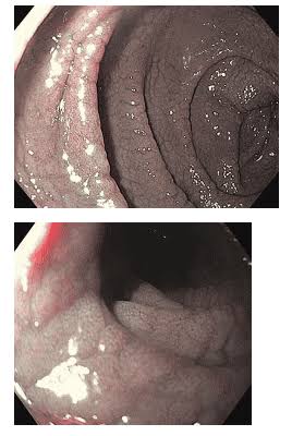
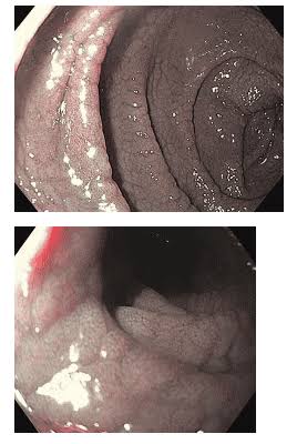
Small intestinal biopsy obtained during upper gastrointestinal endoscopy (gastroscopy) remains the gold standard for confirming celiac disease diagnosis in adults and in children where serology alone is not sufficient for non-biopsy diagnosis. The endoscopist takes multiple biopsies from the second and third parts of the duodenum (at least four biopsies) and from the duodenal bulb (at least one biopsy), as celiac mucosal damage can be patchy — particularly in early or mild disease — and a single inadequate biopsy sample is one of the most common causes of missed or under-diagnosed celiac disease. The biopsies are examined by a histopathologist who classifies the degree of mucosal damage using the Marsh-Oberhuber classification: Marsh 1 shows increased intraepithelial lymphocytes (IELs) above twenty-five per one hundred enterocytes with preserved villous architecture; Marsh 2 adds crypt hyperplasia; Marsh 3a shows partial villous atrophy; Marsh 3b shows subtotal villous atrophy; Marsh 3c shows total villous atrophy — the most severe form. Celiac disease diagnosis requires Marsh 2 or higher changes in the context of positive serology and clinical symptoms compatible with celiac disease. Marsh 1 changes (lymphocytic enteropathy without villous atrophy) are non-specific and can occur in other conditions including H. pylori gastritis, NSAID enteropathy, immune dysregulation, and early celiac disease — requiring careful correlation with serology, HLA status, and clinical context before attributing them to celiac disease. Following diagnosis, repeat biopsy after twelve to twenty-four months of strict gluten-free diet is used to confirm mucosal healing, particularly in patients with persistent symptoms or persistently elevated serology. The degree of mucosal recovery correlates with adherence to the gluten-free diet and is an important indicator of long-term complication risk — unhealed mucosa is associated with continued malabsorption, ongoing nutritional deficiency risk, and (in the long term) elevated risk of rare complications including enteropathy-associated T-cell lymphoma (EATL) and adenocarcinoma of the small bowel, though these complications are rare even in untreated patients. The relationship between malabsorption-related nutritional deficiencies and liver health — including vitamin D deficiency and its association with the bone disease complication of celiac disease, osteoporosis — connects celiac testing to the broader metabolic health picture covered in the article on albumin and liver function.
Celiac Disease Complications and Monitoring Tests
Untreated celiac disease — or celiac disease managed with poor gluten-free diet adherence — is associated with a range of nutritional deficiencies and long-term complications that require specific monitoring tests. The most common nutritional deficiencies in celiac disease include iron deficiency (presenting as anaemia — the most common blood finding leading to celiac disease diagnosis in adults), folate deficiency, vitamin B12 deficiency, vitamin D and calcium deficiency (causing osteoporosis and osteomalacia), zinc deficiency, and magnesium deficiency. Baseline blood tests at celiac disease diagnosis should include a full blood count (for anaemia), serum ferritin and iron studies, folate, vitamin B12, vitamin D, calcium, phosphate, magnesium, and a bone mineral density (DEXA) scan in adults with established disease to assess for osteoporosis. These deficiencies arise from the malabsorptive state caused by villous atrophy in the proximal small bowel — the region where iron, folate, vitamin D, and calcium are preferentially absorbed. On a strict gluten-free diet with mucosal healing, absorption normalises and nutritional deficiencies correct — though supplementation is often needed in the short term while the mucosa recovers. Annual blood tests for known celiac disease patients typically include anti-tTG IgA (as a dietary adherence surrogate), full blood count, ferritin, vitamin D, and B12 — escalating to repeat biopsy if serology remains elevated or symptoms persist. Refractory celiac disease — where symptoms and villous atrophy persist despite strict gluten-free diet adherence — requires specialist gastroenterology assessment to exclude complications including refractory sprue type I and type II, the latter of which carries lymphoma risk and warrants specialist management. The liver enzyme pattern in untreated celiac disease often includes mild transaminase elevation (ALT and AST) that resolves on a gluten-free diet — the connection between celiac disease and liver health is explored further in the article on ALT and AST blood tests.
When to Get Tested and Who Should Be Screened
Celiac disease testing is indicated across a broad range of clinical presentations — not only in patients with classic gastrointestinal symptoms of diarrhoea, bloating, and weight loss, but in the many situations where celiac disease may be the underlying diagnosis driving an apparently unrelated clinical finding. Current guidelines recommend celiac testing in: unexplained iron deficiency anaemia (particularly in premenopausal women or children where iron loss cannot explain the degree of anaemia); unexplained osteoporosis or low bone density particularly in young adults; recurrent miscarriage or unexplained infertility; first-degree relatives of diagnosed celiac disease patients (prevalence is approximately ten percent — ten times the general population); patients with type 1 diabetes mellitus (co-prevalence approximately six to ten percent given shared HLA genetics); patients with autoimmune thyroid disease; unexplained peripheral neuropathy or cerebellar ataxia (neurological celiac disease — gluten ataxia — is a distinct and important presentation); dermatitis herpetiformis (the skin manifestation of celiac disease, characterised by intensely itchy vesicular rash on extensor surfaces, which requires separate skin biopsy for direct immunofluorescence diagnosis but should be managed with the same gluten-free diet); and patients with unexplained mild transaminase elevation where common causes have been excluded. Screening of entirely asymptomatic individuals without risk factors or associated conditions is not currently recommended outside of research settings. However, the low threshold for testing across these diverse clinical scenarios reflects the reality that celiac disease is chronically underdiagnosed, that the morbidity of undiagnosed celiac disease accumulates over years, and that the treatment — a strict gluten-free diet — while requiring significant lifestyle adjustment is highly effective at reversing the morbidity of the condition when adhered to. The article on when digestive symptoms need medical attention covers the specific symptom threshold at which investigation including celiac disease testing should be sought.
Is a gluten-free diet safe to start without formal diagnosis?
Starting a gluten-free diet without formal diagnosis is strongly discouraged by gastroenterology guidelines and for good reason. As described above, self-initiated gluten avoidance before appropriate testing renders the diagnostic tests inaccurate — a patient who feels better on a gluten-free diet cannot then have accurate celiac testing without reintroducing gluten for six weeks, which many find difficult after experiencing symptom improvement. This creates a scenario where patients commit to a lifelong, expensive, and socially demanding diet without a confirmed diagnosis — and without knowing whether their condition is celiac disease (requiring strict lifelong avoidance to prevent complications), non-celiac gluten sensitivity (a distinct condition with less stringent requirements and no autoimmune or malabsorptive complications), wheat allergy, or an entirely unrelated condition where gluten avoidance produced a placebo-driven symptom improvement. Formal diagnosis matters: it determines whether you need to completely avoid cross-contamination (celiac disease), whether family members need screening (celiac disease — a genetic condition with ten percent first-degree relative prevalence), whether you need long-term nutritional monitoring and bone density assessment, and whether your diet needs to be managed by a specialist dietitian with coeliac disease expertise. If you are experiencing symptoms that make you suspect gluten intolerance, the right approach is to discuss testing with your GP before modifying your diet — or, if you have already started a gluten-free diet, to discuss the gluten reintroduction protocol and HLA testing with your gastroenterologist to clarify the diagnosis. The diagnostic framework for celiac disease, combined with the full picture of gastrointestinal and liver health testing covered across this site — from liver function tests to GGT — provides the evidence base for making informed decisions about digestive health investigation.
I wish I had read this article before I started a gluten-free diet on my own. I felt better almost immediately and assumed I must have celiac disease — but when I went to my GP for blood tests six months later, everything came back normal. My gastroenterologist explained that because I’d already been gluten-free for six months, the antibodies had fallen and the diagnosis couldn’t be confirmed without a gluten challenge. I’m now three weeks into reintroducing gluten and it has been really difficult. Please get tested before you try the diet.
Emma, your experience is unfortunately very common — the diet-before-diagnosis scenario is one of the most frequent diagnostic challenges in celiac disease. The good news is that the gluten challenge, while difficult, is the right path to a confirmed diagnosis that will serve you well for the rest of your life. Knowing whether you have true celiac disease (requiring strict lifelong avoidance) versus non-celiac gluten sensitivity (where occasional exposure is less harmful) matters enormously for your long-term management, family screening decisions, and monitoring plan. Hang in there through the challenge — the answer is worth it.
It took eight years from my first symptoms to my celiac diagnosis. I had iron deficiency anaemia for years that kept being attributed to ‘poor diet’, and my GP never once considered celiac as the cause. Eventually a hospital dietitian suggested celiac testing after I was admitted for severe anaemia — anti-tTG IgA came back at 15 times the upper limit of normal. The bone density scan showed significant osteopenia at age 34 that could have been prevented with earlier diagnosis.