
Stool tests — laboratory examinations of faecal samples — are among the most valuable and yet most underutilised diagnostic tools in digestive health. They can detect blood hidden in the stool before it is visible to the naked eye, identify bacterial, viral, and parasitic infections causing gastroenteritis, diagnose inflammatory bowel disease activity through biomarkers, test for colorectal cancer without colonoscopy, identify specific pathogens causing diarrhoea, and screen for Helicobacter pylori and celiac disease. Understanding what different stool tests measure, what they are used for, and how to interpret their results puts patients in a better position to understand why their doctor has ordered a particular stool test and what the findings mean for their digestive health management.
The range of stool tests available spans from simple, inexpensive bedside tests (faecal occult blood testing by guaiac card) to complex molecular platforms (multiplex PCR gastrointestinal pathogen panels detecting dozens of pathogens simultaneously from a single sample). Each test is designed to answer a specific clinical question, and understanding which question each test is built to answer is the foundation of appropriate test selection and result interpretation. A test that is excellent for colorectal cancer screening is not appropriate for diagnosing infectious diarrhoea; a test that identifies acute bacterial infection does not evaluate chronic inflammation. This article covers the principal stool tests used in clinical practice, their clinical applications, what results mean, and the practical considerations patients need to be aware of when providing stool samples for testing.
Faecal Occult Blood Tests (FOBT and FIT)
Faecal occult blood testing detects blood in the stool that is not visible to the naked eye — the “occult” (hidden) blood that can indicate colorectal cancer or advanced polyps bleeding into the gastrointestinal lumen at quantities too small to change stool colour. Two main types of faecal occult blood tests are used in clinical practice: the guaiac-based FOBT (gFOBT) and the faecal immunochemical test (FIT, also called iFOBT). Guaiac FOBT (gFOBT) detects the peroxidase activity of haemoglobin by a chemical colour reaction. Because it reacts to haemoglobin from any source — human or dietary — dietary restrictions are required before the test (red meat, certain vegetables like broccoli and turnips containing plant peroxidases, and aspirin must be avoided for two to three days before testing to prevent false positives). gFOBT has been largely superseded by FIT in modern colorectal cancer screening programmes but remains in use in some settings. Faecal immunochemical test (FIT) uses antibodies specific to human haemoglobin, making it specific to human lower gastrointestinal bleeding rather than dietary haemoglobin or upper GI bleeding (haemoglobin from upper GI sources is digested before reaching the lower colon where FIT samples). FIT requires no dietary restrictions, is more specific for colorectal cancer than gFOBT, and has replaced gFOBT in the US Colorectal Cancer Screening guidelines and the NHS bowel screening programme. A positive FIT result requires colonoscopy follow-up to investigate the source of the occult blood. The article on when digestive symptoms require medical attention covers the symptom context in which stool testing is most urgently needed.
Faecal Calprotectin — Intestinal Inflammation Marker
Faecal calprotectin is a protein released by neutrophils (white blood cells) as they migrate through inflamed intestinal mucosa into the gastrointestinal lumen, making it a highly sensitive marker of intestinal inflammation. The stool calprotectin test measures the concentration of this protein in a faecal sample, with results reported in μg/g of stool. Normal calprotectin is generally below 50 μg/g; values between 50 and 200 μg/g are borderline and often require clinical correlation or repeat testing; values above 200 μg/g indicate significant intestinal inflammation requiring further investigation. The test’s primary clinical utility is in distinguishing inflammatory bowel disease (IBD — Crohn’s disease or ulcerative colitis) from irritable bowel syndrome (IBS) in patients presenting with chronic abdominal pain, diarrhoea, and bloating. This distinction is clinically critical because IBD and IBS present with similar symptoms but require completely different management: IBD requires endoscopic assessment, mucosal biopsy, and disease-modifying therapy, while IBS is managed with dietary and lifestyle modification, psychological support, and symptom-targeted pharmacotherapy. A normal calprotectin (below 50 μg/g) in a symptomatic patient makes active IBD very unlikely — sensitivity exceeds ninety percent — and can safely redirect the diagnostic pathway away from colonoscopy in primary care, potentially avoiding unnecessary invasive investigation.
Beyond initial diagnosis, faecal calprotectin is an established monitoring tool for IBD activity in patients with known Crohn’s disease or ulcerative colitis. Serial calprotectin measurement correlates with endoscopic mucosal inflammation — a rising calprotectin in a patient on IBD treatment may precede a clinical flare by weeks to months, providing an early warning that treatment is losing effectiveness and that therapy escalation or treatment switch should be considered before the patient deteriorates significantly. Calprotectin above 250 μg/g in a patient with known IBD is generally associated with endoscopically active disease, while values below 100 μg/g are associated with mucosal healing. This serial monitoring role makes stool calprotectin a valuable non-invasive surrogate for repeat colonoscopy in IBD management — though it does not replace endoscopy when tissue diagnosis, disease extent mapping, or dysplasia surveillance is required. The relationship between bowel inflammation markers and the visible changes in stool that patients notice — including the pale stools covered in the article on pale stool and bile duct problems — illustrates how laboratory and clinical findings complement each other in digestive health assessment.
Stool Cultures and Molecular GI Pathogen Panels
Stool culture — the traditional test for identifying bacterial causes of infectious gastroenteritis — involves plating faecal samples on selective growth media to identify pathogens including Salmonella, Shigella, Campylobacter, Shiga toxin-producing E. coli (STEC, including O157:H7), and Yersinia. Culture remains the gold standard for isolating viable bacteria, identifying antibiotic susceptibility patterns, and performing public health strain typing for outbreak investigations. However, stool culture has practical limitations: it takes two to four days to produce results, sensitivity varies by organism (Campylobacter requires specific culture conditions), and culture cannot detect viral or parasitic pathogens. Most bacterial gastroenteritis in otherwise healthy adults in developed countries is self-limiting and does not require antibiotic treatment — stool culture is most indicated in patients with severe symptoms (high fever, bloody diarrhoea, signs of dehydration or systemic sepsis), healthcare workers, food handlers, and those with immunocompromise. Testing for Clostridioides difficile (C. diff) — the toxin-producing bacterium causing antibiotic-associated diarrhoea — is performed separately using stool glutamate dehydrogenase (GDH) antigen and toxin A/B immunoassay, or PCR for toxin genes; it should be ordered in patients with diarrhoea following antibiotic use or hospitalisation rather than as part of routine gastroenteritis workup.
Molecular gastrointestinal pathogen panels — multiplex PCR platforms such as the BioFire GI Panel — can simultaneously detect dozens of bacterial, viral, and parasitic pathogens from a single stool sample within one to two hours, dramatically expanding diagnostic capability compared to traditional culture. These panels typically detect common bacteria (Salmonella, Campylobacter, Shigella, E. coli species, C. diff), viruses (norovirus, rotavirus, adenovirus, sapovirus), and parasites (Giardia, Cryptosporidium, Cyclospora, Entamoeba histolytica). Molecular panels are particularly useful in immunocompromised patients (where a broader range of pathogens can cause disease), in patients with persistent diarrhoea unresponsive to empirical treatment, in travellers returning from endemic regions, and during community or healthcare outbreaks where rapid pathogen identification guides public health response. A potential limitation is that molecular panels detect genetic material of pathogens even after clinical resolution of infection, so a positive PCR result requires interpretation alongside clinical symptoms and the timing of testing relative to symptom onset. The article on dark urine and its significance addresses how some gastrointestinal infections — particularly hepatitis A — can produce liver involvement alongside diarrhoeal symptoms, making integrated clinical assessment important.
Faecal Elastase and Pancreatic Function Testing
Faecal elastase-1 is an enzyme secreted by the pancreas into the small intestine that remains stable through transit to the colon and can be measured in a random stool sample to assess exocrine pancreatic function. Low faecal elastase-1 (below 100 μg/g) indicates severe exocrine pancreatic insufficiency (EPI) — the condition where the pancreas fails to produce adequate digestive enzymes, producing malabsorption of fats, proteins, and fat-soluble vitamins (A, D, E, K). Values between 100 and 200 μg/g indicate moderate insufficiency; values above 200 μg/g are generally considered normal. EPI causes steatorrhoea — pale, greasy, foul-smelling stools that float due to unabsorbed fat — alongside weight loss, nutritional deficiencies, and diabetes from concurrent endocrine pancreatic dysfunction. Causes include chronic pancreatitis (the most common cause), pancreatic surgery, cystic fibrosis, and pancreatic cancer. Treatment with pancreatic enzyme replacement therapy (PERT — enteric-coated pancreatic enzyme capsules taken with meals) is highly effective at controlling steatorrhoea and preventing nutritional deficiencies when EPI is identified and treated. Stool elastase testing should be considered in any patient with unexplained steatorrhoea, weight loss, or fat-soluble vitamin deficiencies, particularly in the context of a history of alcohol use (chronic pancreatitis risk), previous pancreatic surgery, or cystic fibrosis. The pale, greasy stool appearance of severe fat malabsorption is related to but distinct from the pale stool of biliary obstruction — context and clinical history differentiate these effectively.
Frequently Asked Questions About Stool Tests
How do I collect a stool sample correctly?
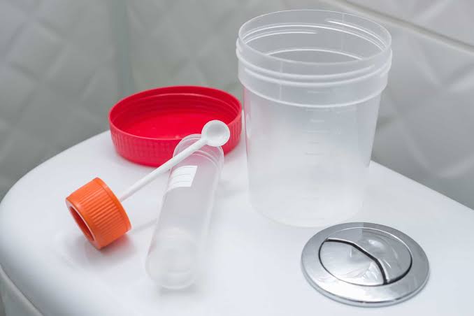
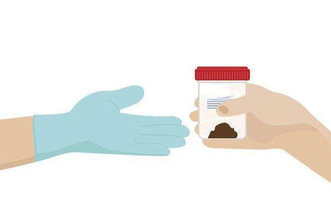
Correct stool sample collection ensures that test results are accurate and avoids the need for repeat sampling. Most stool tests require a fresh sample — typically collected within one to two hours of production and placed in the laboratory-provided collection container using the spatula or spoon attached to the container lid. The sample should not be contaminated with toilet water, urine, or toilet paper, which can degrade the sample or interfere with testing. Some laboratories provide collection aids (a cardboard sheet placed across the toilet bowl) to make collection easier without water contact. For FIT (faecal immunochemical test), the sample must not be collected during or within three days of menstruation, from patients with known haemorrhoids actively bleeding, or from patients with visible blood in the stool already — as these sources of blood will produce a positive result that does not reflect the colorectal cancer screening intent of the test. For calprotectin testing, the sample should ideally not be collected during an acute non-IBD gastrointestinal infection (viral gastroenteritis, food poisoning) as these will transiently elevate calprotectin through non-IBD mucosal inflammation and could lead to false-positive interpretation. Most samples should be refrigerated and transported to the laboratory within twenty-four to forty-eight hours.
My FIT test came back positive — what happens next?
A positive FIT result means that blood was detected in the stool at a level above the test’s threshold — it does not mean you have cancer. The majority of positive FIT results are caused by benign sources of occult bleeding including haemorrhoids, anal fissures, benign colorectal polyps, diverticular disease, and non-steroidal anti-inflammatory drug (NSAID) use causing mucosal irritation. However, the reason a positive FIT requires colonoscopy follow-up is that it can also indicate advanced polyps (pre-cancerous lesions) or colorectal cancer, and distinguishing these from benign causes requires direct visualisation of the colon by colonoscopy. The positive predictive value of a single positive FIT for colorectal cancer is approximately five to ten percent — so the majority of patients with a positive FIT will have normal colonoscopy or benign findings, but the consequences of missing the minority with significant pathology make colonoscopy investigation mandatory. Colonoscopy should ideally be performed within eight to twelve weeks of a positive FIT result, in line with national colorectal cancer screening programme guidelines. The article on fatigue and its association with bowel and liver disease addresses symptoms that often accompany colorectal pathology and the importance of early evaluation.
Sources: USPSTF — Colorectal Cancer Screening · ACG — Stool Tests for Colon Cancer · NIDDK — Digestive Diseases
H. pylori Stool Antigen Test
The Helicobacter pylori stool antigen test (HpSA) detects H. pylori antigens — fragments of the bacterium’s proteins — in a faecal sample using monoclonal antibody-based immunoassay. H. pylori is a bacterial infection of the gastric mucosa affecting approximately forty-four percent of the global population and is the leading cause of peptic ulcer disease, chronic active gastritis, and gastric cancer. The stool antigen test is one of three preferred non-invasive tests for H. pylori detection alongside the urea breath test (UBT) and serological antibody testing. Unlike serology — which detects IgG antibodies that persist for months to years after eradication and cannot confirm active infection — the stool antigen test and urea breath test both detect active, current infection and can confirm successful eradication after treatment. The HpSA test has sensitivity and specificity exceeding ninety percent for detecting active H. pylori infection and is the preferred non-invasive H. pylori test in many settings because it requires only a stool sample rather than a clinical procedure. For accurate results, proton pump inhibitors (PPIs) must be stopped two weeks before testing and antibiotics four weeks before — as both suppress H. pylori below detectable thresholds without eradicating it, producing false-negative results. The stool antigen test is appropriate for initial H. pylori diagnosis in patients with dyspepsia without alarm symptoms, for post-treatment eradication confirmation (tested at least four weeks after completing antibiotic therapy), and for monitoring in populations with high gastric cancer risk. The article on hepatitis blood tests addresses the blood-based testing complement to the stool-based diagnostic toolkit in gastrointestinal infection workup.
Stool Ova and Parasite Examination
The stool ova and parasite (O&P) examination — microscopic examination of faecal samples for parasitic organisms and their eggs (ova) — remains the traditional method for identifying intestinal parasites causing diarrhoea, abdominal pain, and malabsorption. The examination typically requires three stool samples collected on three separate days (because parasite shedding is intermittent and a single sample may miss infection), concentrated and examined by a trained microscopist for trophozoites, cysts, oocysts, and ova. Organisms commonly detected by O&P examination include Giardia lamblia (giardiasis — the most common intestinal protozoal infection in developed countries, causing persistent watery diarrhoea, bloating, and fat malabsorption after ingestion of contaminated water or food), Entamoeba histolytica (amoebiasis — causes invasive colitis and liver abscess in endemic regions), Cryptosporidium parvum (causes severe watery diarrhoea in immunocompromised patients, particularly HIV/AIDS and transplant recipients), Strongyloides stercoralis (roundworm capable of causing hyperinfection syndrome in immunosuppressed patients), and tapeworm segments (Taenia spp.). O&P examination is most clinically indicated in patients with persistent diarrhoea lasting more than fourteen days, recent travel to endemic regions, immunocompromise, occupation with animal exposure, or exposure to contaminated water sources. Sensitivity of traditional microscopy varies by organism and laboratory expertise — molecular panel detection of Giardia and Cryptosporidium is now preferred in many centres given superior sensitivity. Stool O&P examination remains important for detecting organisms not included in molecular panels, for characterising tapeworm species, and in resource-limited settings where molecular testing is unavailable. Treatment is organism-specific: Giardia responds to metronidazole or tinidazole; Cryptosporidium in immunocompetent patients is usually self-limiting while immunocompromised patients require nitazoxanide and immune reconstitution where possible; Strongyloides requires ivermectin. The significance of gastrointestinal infection burden on systemic health — including the hepatic effects of amoebiasis producing liver abscess — connects stool-based infection testing to broader diagnostics including liver function test panels used to evaluate extraluminal infection spread.
Interpreting Stool Test Results in Context
No stool test result should be interpreted in isolation — each result must be contextualised against the patient’s symptoms, clinical history, medication list, travel and dietary history, and the pre-test probability of the condition being tested for. A positive calprotectin in a patient with acute viral gastroenteritis does not indicate IBD; a negative FIT does not exclude colorectal cancer in a patient with alarm symptoms (rectal bleeding, weight loss, change in bowel habit) who requires colonoscopy on clinical grounds regardless of FIT result; a negative stool culture in a patient with severe bloody diarrhoea and fever does not exclude bacterial infection when the sample was collected after antibiotic initiation or when the responsible organism requires specific culture conditions. The guiding principle of stool test interpretation is that a negative test result reduces the probability of a disease, while a positive result increases it — but neither a positive nor negative result is the end of the diagnostic process in the majority of cases. A patient with classic IBD symptoms, elevated calprotectin, and normal blood tests still requires colonoscopy with mucosal biopsy to confirm IBD diagnosis, characterise disease extent, and obtain histopathological confirmation before committing to long-term immunosuppressive therapy. Understanding the role of stool tests within the broader digestive health investigation framework — alongside blood tests, imaging, and endoscopy — allows patients and clinicians to make appropriate decisions about which investigations to pursue, in what order, and what thresholds should prompt escalation of investigation. The liver and digestive health diagnostic ecosystem, covered across multiple articles on this site including those on ALT and AST, bilirubin testing, and the GGT test, reflects the reality that no single test answers every clinical question — thoughtful test selection and result integration are the hallmarks of accurate digestive disease diagnosis.
Can I use stool tests to monitor colorectal cancer treatment?
While the FIT and gFOBT are primarily screening tests used to detect early-stage colorectal cancer or pre-cancerous polyps before diagnosis, stool tests are not typically used to monitor treatment response in patients already diagnosed with colorectal cancer — blood-based tumour marker monitoring (carcinoembryonic antigen, CEA) and imaging surveillance are the standard post-treatment monitoring tools for colorectal cancer. However, stool tests retain a role in post-polypectomy surveillance in specific circumstances — patients who had a FIT-positive colonoscopy that found and removed only hyperplastic polyps may be re-enrolled in the standard FIT screening programme rather than receiving colonoscopy-based surveillance. Patients with inflammatory bowel disease who have been treated for colorectal dysplasia require ongoing colonoscopy-based surveillance, and faecal calprotectin monitoring for IBD activity continues alongside cancer surveillance. The integration of stool-based testing into the broader cancer screening ecosystem reflects the role of non-invasive tests in population health: making high-compliance, low-burden screening available to the largest possible number of people to detect disease at a stage where curative treatment is still achievable. For patients with liver or biliary involvement from colorectal cancer metastases, the liver function tests covered in the liver function test overview and alkaline phosphatase testing become part of the monitoring framework alongside stool and imaging surveillance.
Stool testing is a practical, non-invasive first-line approach in digestive health assessment — a tool that helps clinicians triage patients appropriately, identifies conditions requiring urgent investigation, and monitors known diseases over time without the burden and risk of endoscopic procedures. Understanding what to expect from stool tests, how to collect samples correctly, and what results mean empowers patients to engage actively and effectively in their digestive health care — from cancer screening participation to IBD monitoring to prompt post-treatment H. pylori confirmation. The breadth of what stool tests can reveal, from infection to inflammation to cancer and pancreatic insufficiency, makes them an essential component of the digestive health diagnostic toolkit.
My GP ordered a calprotectin test when I went in with three months of bloating and loose stools. I expected her to just say it was IBS but she said she wanted to rule out IBD first. The result came back at 320 and I ended up having a colonoscopy that diagnosed Crohn’s disease. I’m now on medication and feeling so much better. I had no idea a simple stool test could lead to such an important diagnosis.
Sandra, your experience illustrates exactly why calprotectin has become such a valuable primary care test — it can identify patients with active intestinal inflammation who need colonoscopy rather than reassurance, even when symptoms alone could fit IBS. A calprotectin of 320 μg/g is clearly in the range associated with active IBD, and it’s reassuring that your GP acted on it promptly. Early diagnosis in Crohn’s disease, before significant bowel damage has occurred, gives the best chance of achieving and maintaining remission with medical therapy.
I want to mention that the FIT test through the NHS bowel screening programme genuinely saved my father’s life — he had no symptoms at all, the FIT was positive, colonoscopy found a stage 1 colorectal cancer that was removed successfully. He’s been cancer free for five years. Everyone over 50 should take these tests seriously when they arrive in the post.