
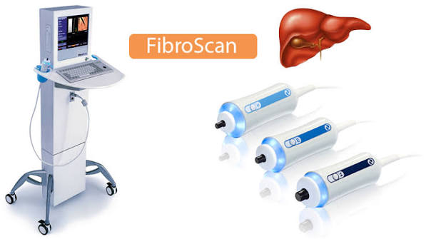
FibroScan is a non-invasive medical device that measures liver stiffness and liver fat content using a technology called transient elastography and controlled attenuation parameter (CAP). Developed by Echosens and approved by the FDA and CE Mark, FibroScan has transformed the non-invasive assessment of liver fibrosis — the degree of scarring in the liver — and is now a standard tool in liver clinics worldwide. For patients with fatty liver disease (MASLD/NAFLD), chronic viral hepatitis, alcoholic liver disease, or other chronic liver conditions, FibroScan provides a rapid, painless, bedside assessment of liver health that avoids the need for liver biopsy in the majority of patients. Understanding what FibroScan measures, how it works, how to interpret the results (liver stiffness in kPa and CAP score in dB/m), and when it should be repeated provides patients with the knowledge to engage actively with their liver disease monitoring and management.
Before FibroScan and similar non-invasive fibrosis assessment tools became available, the only reliable method for quantifying the degree of liver fibrosis was percutaneous liver biopsy — a procedure involving insertion of a needle through the skin into the liver to obtain a small core of tissue for histopathological analysis. While liver biopsy remains the gold standard for fibrosis diagnosis (and is still required in complex cases where non-invasive results are discordant), it carries a small but real risk of complications (bleeding in approximately 0.3–0.5% of procedures, pain, and rarely organ perforation), produces a sample representing only approximately one fifty-thousandth of the total liver volume (sampling error), and cannot be repeated frequently for monitoring. FibroScan addresses these limitations by providing a non-invasive, rapid (five to ten minutes), repeatable assessment that can be performed in the clinic setting without preparation or recovery time. Its clinical validation across tens of thousands of patients has established robust diagnostic thresholds for different stages of fibrosis in different liver disease aetiologies, making it an evidence-based alternative to biopsy for the majority of clinical questions in chronic liver disease management.
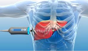
How FibroScan Works: Transient Elastography Explained
FibroScan uses transient elastography — a technique that measures the propagation speed of a low-frequency mechanical shear wave through the liver tissue. A specialised probe is placed on the skin in the intercostal space (between two ribs) in the right upper quadrant, and a brief vibration is generated that produces a shear wave propagating through the underlying liver tissue. A simultaneous ultrasound pulse tracks the shear wave’s propagation speed. Because liver stiffness increases as fibrosis accumulates (fibrotic scar tissue is significantly stiffer than normal hepatocytes), the shear wave travels faster through a fibrotic liver than through a normal one. The FibroScan device converts shear wave propagation speed into liver stiffness, reported in kilopascals (kPa). A valid FibroScan result requires at least ten valid measurements with an interquartile range (IQR) of less than thirty percent of the median value — ensuring measurement consistency and reliability. The Controlled Attenuation Parameter (CAP) is measured simultaneously by the FibroScan device: it uses the ultrasound signal attenuation (reduction in ultrasound wave amplitude with depth) to estimate the degree of hepatic steatosis (fat content). Because fat attenuates ultrasound more than normal liver parenchyma, higher CAP values indicate greater fat content. CAP is reported in decibels per metre (dB/m); values below 238 dB/m are generally considered normal (S0); 238–258 dB/m indicates mild steatosis (S1); 259–290 dB/m indicates moderate steatosis (S2); above 290 dB/m indicates severe steatosis (S3). Together, liver stiffness and CAP provide a rapid, dual-parameter non-invasive assessment of both the degree of fibrosis and the degree of fatty infiltration — the two most clinically important structural parameters in chronic liver disease monitoring.
Interpreting FibroScan Results: Liver Stiffness Values
Liver stiffness values from FibroScan are interpreted using disease-specific thresholds, because the normal liver stiffness and the stiffness values associated with significant fibrosis differ between disease aetiologies (due to differences in inflammation, cholestasis, and congestion that can affect liver stiffness independently of fibrosis). The following thresholds are commonly used as reference guides — noting that exact cut-offs vary between different published guidelines and should always be interpreted in clinical context: For MASLD/NAFLD (metabolic fatty liver disease): below 7.0 kPa is consistent with no significant fibrosis (F0–F1); 7.0–9.5 kPa suggests significant fibrosis (F2); 9.5–12 kPa suggests advanced fibrosis (F3); above 12–13 kPa suggests cirrhosis (F4). For chronic hepatitis C: below 7.1 kPa is consistent with F0–F1; 7.1–9.5 kPa for F2; 9.5–12.5 kPa for F3; above 12.5 kPa for F4 (cirrhosis). For chronic hepatitis B: below 6.0 kPa suggests F0–F1; 6.0–8.0 kPa for F2; 8.0–11 kPa for F3; above 11 kPa for cirrhosis. For alcoholic liver disease: cut-offs are shifted higher because alcohol causes hepatic inflammation and congestion that elevate liver stiffness independently of fibrosis. Important confounders of liver stiffness that must be considered when interpreting results: acute hepatitis (severe hepatocellular inflammation elevates liver stiffness regardless of fibrosis stage); active alcohol use (even without inflammation, acute alcohol use within twenty-four hours elevates liver stiffness); bile duct obstruction (cholestasis stiffens the liver); post-prandial state (eating increases splanchnic blood flow and portal pressure, elevating liver stiffness — FibroScan should be performed fasting for at least two hours for accurate results); right heart failure (elevated right atrial pressure increases hepatic venous pressure and liver stiffness). The blood test pattern that accompanies elevated liver stiffness — elevated ALT, AST, and GGT — is covered in the articles on ALT and AST blood tests and GGT testing.
When FibroScan Is Used and Its Limitations
FibroScan is indicated in the following clinical contexts: Initial fibrosis assessment in patients with newly diagnosed chronic liver disease (MASLD, chronic viral hepatitis, alcoholic liver disease, autoimmune liver disease) to determine baseline fibrosis stage and guide management intensity. Patients with advanced fibrosis (F3–F4) require more intensive monitoring, HCC surveillance, and consideration of treatment to prevent progression. Monitoring fibrosis progression in patients with known chronic liver disease — serial FibroScan measurements (typically every one to two years) track whether fibrosis is stable, progressing, or regressing with treatment or lifestyle intervention. Successful HCV treatment with SVR (viral cure) leads to liver stiffness reduction over twelve to eighteen months in most patients. Weight loss in MASLD leads to measurable CAP and stiffness reduction. Post-treatment response assessment — after antiviral treatment for hepatitis C or B, FibroScan confirms fibrosis regression and determines whether HCC surveillance protocols can be modified based on reduced liver stiffness. Avoiding liver biopsy in patients with concordant non-invasive fibrosis assessment — when FibroScan result is consistent with clinical history, blood tests, and imaging, biopsy can be avoided in the majority of cases; biopsy is reserved for discordant results, suspected multiple coexisting liver diseases, or when histological assessment of disease activity (inflammation grade) is specifically required. Technical limitations of FibroScan include: failure rate in obese patients — standard M probe has reduced accuracy in patients with a skin-to-liver capsule distance above 25 mm (typically BMI above 28–30), where the XL probe (designed for obese patients) is required and not available in all centres; ascites — the presence of peritoneal fluid between the skin and liver prevents shear wave transmission, rendering FibroScan unreliable in patients with significant ascites; narrow intercostal spaces in some patients. When FibroScan is technically unreliable, MR elastography (as discussed in the dedicated MRI article) provides the most accurate alternative. The clinical utility of FibroScan in the context of the full liver investigation pathway — including ultrasound and liver blood tests — is described in the article on liver ultrasound.
Frequently Asked Questions About FibroScan
My FibroScan showed 8.2 kPa — what does this mean for me?
A liver stiffness of 8.2 kPa places most patients in the range consistent with significant fibrosis (F2) for MASLD or hepatitis C aetiology, or potentially at the lower end of F3 for hepatitis B — the exact interpretation depends on which liver disease is present. This result means there is evidence of meaningful liver scarring beyond the minimal fibrosis of early disease, but is not in the cirrhosis range (which typically begins at 12–13 kPa for MASLD). The appropriate response to an 8.2 kPa result depends on the clinical context: in a patient with MASLD and this stiffness, the priority is addressing the modifiable risk factors (weight loss, metabolic syndrome control, alcohol cessation) and monitoring with repeat FibroScan in one to two years; in a patient with chronic hepatitis C at this stiffness, antiviral treatment with DAAs should be prioritised to prevent progression to advanced fibrosis; in chronic hepatitis B, antiviral treatment eligibility depends on this stiffness alongside viral load and ALT. In all cases, a FibroScan result should prompt a consultation with your GP or hepatologist to interpret the result in the context of your specific liver condition, blood test results, and risk factors. The relationship between liver stiffness and the symptoms of advanced liver disease — including the fatigue covered in the article on fatigue and liver health — helps contextualise what this result means for your overall health.
How do I prepare for a FibroScan?
FibroScan preparation is simple — the key requirement is fasting for at least two to three hours before the test (food and alcohol both elevate liver stiffness through increased portal blood flow and hepatic venous congestion respectively). Water is permitted. Patients should avoid alcohol for twenty-four hours before the test to eliminate alcohol-related liver stiffness elevation. The test itself takes five to ten minutes in a clinic setting. The sonographer or nurse applies ultrasound gel to the right side of the abdomen, places the probe between the ribs, and asks the patient to lie still and breathe normally while measurements are taken. Ten measurements are required for a valid result. There is no radiation, no needles, no fasting beyond two to three hours, and no recovery time — patients can drive and return to normal activities immediately after the test. FibroScan results are available immediately and are typically discussed with the patient or their clinician on the same day or at a follow-up appointment. Annual FibroScan monitoring is recommended in patients with known significant fibrosis (F2 and above), with more frequent testing in patients with active disease progression risk factors. The broader context of what liver investigations can tell you — from blood tests through imaging to non-invasive fibrosis assessment — is covered across the liver and digestive health series on this site, including the comprehensive overview of liver function tests and the imaging articles on liver ultrasound and abdominal ultrasound.
Sources: EASL — Non-Invasive Tests for Liver Disease · AASLD — Liver Disease Guidance · NIDDK — NAFLD and NASH
FibroScan vs Liver Biopsy: Understanding the Trade-offs
The question patients and clinicians face when assessing liver fibrosis is not simply whether to use FibroScan or liver biopsy — it is how to select the most appropriate investigation for the specific clinical question, balancing diagnostic accuracy, patient safety, practicality, and cost. FibroScan and liver biopsy provide complementary rather than competing information, and understanding their respective strengths and limitations helps patients understand why one or the other (or both) may be recommended in specific circumstances.
Liver biopsy advantages: Biopsy remains the only investigation that can simultaneously assess fibrosis stage, inflammatory activity (necroinflammatory grade), steatosis percentage, cellular ballooning, glycogenated nuclei, Mallory-Denk bodies, and other histological features that define disease activity and predict progression independently of fibrosis. In MASLD, the NAFLD Activity Score (NAS) derived from biopsy — combining steatosis, lobular inflammation, and hepatocellular ballooning — is the gold standard for disease activity assessment and remains essential for clinical trial endpoints. In autoimmune hepatitis, biopsy assesses interface hepatitis activity that guides corticosteroid treatment decisions. When two coexisting liver diseases are suspected (e.g., MASLD plus alcoholic liver disease, or hepatitis B plus autoimmune hepatitis), biopsy can assess both simultaneously, while FibroScan reflects only the combined structural consequence.
FibroScan advantages: FibroScan is performed without needles, anaesthesia, or recovery time, and can be repeated as frequently as clinically needed (annually or more often) for longitudinal fibrosis monitoring — biopsy sampling error, procedure risk, and patient acceptance limit biopsy to a small number of lifetime assessments. FibroScan is available as a point-of-care clinic tool, whereas biopsy requires a procedure suite, consent, post-procedure monitoring, and pathology reporting with a one to two week turnaround. FibroScan is considerably more cost-effective for monitoring stable patients. The clinical consensus — reflected in EASL (European Association for the Study of the Liver) and AASLD (American Association for the Study of Liver Diseases) guidelines — is that FibroScan and other validated non-invasive tests should be the first-line method for fibrosis assessment in chronic liver disease, with biopsy reserved for discordant or equivocal non-invasive results, suspected overlap syndromes, and clinical trial enrolment. The relationship between the blood tests that initially direct patients towards liver fibrosis assessment — including ALT and AST, GGT, and albumin — and the FibroScan that quantifies the structural consequence of liver injury provides the key non-invasive diagnostic framework used in modern liver clinics.
FibroScan in MASLD and Metabolic Liver Disease
Metabolic dysfunction-associated steatotic liver disease (MASLD — the renamed and redefined entity previously called NAFLD) is the most common liver disease in Western populations, affecting an estimated twenty-five percent of adults globally. FibroScan has become the central non-invasive monitoring tool in MASLD management because it simultaneously quantifies both the degree of fat accumulation (via CAP) and the degree of fibrosis (via liver stiffness), tracking the two most clinically important parameters of MASLD progression in a single five-minute test.
In MASLD, fibrosis stage is the strongest independent predictor of liver-related mortality and overall survival — more so than the degree of steatosis or inflammation alone. Patients with MASLD and advanced fibrosis (F3–F4) have a substantially higher risk of liver-related complications (decompensated cirrhosis, hepatocellular carcinoma, liver transplantation) and all-cause mortality compared to those with F0–F2 fibrosis. The identification of advanced fibrosis therefore directly drives clinical management intensity: patients with F3–F4 fibrosis require six-monthly HCC surveillance ultrasound (as covered in the liver ultrasound article), upper GI endoscopy for varices screening, hepatology clinic follow-up, and consideration of emerging MASLD-specific pharmacological treatments (the FXR agonist obeticholic acid and newer PPAR agonists that have demonstrated fibrosis regression in phase 3 trials). Patients with F0–F2 fibrosis are managed primarily through lifestyle modification with annual FibroScan monitoring.
The CAP score’s utility in MASLD extends beyond simple steatosis quantification. Reduction in CAP score following a dietary intervention or weight loss programme provides objective evidence of hepatic steatosis regression that validates the effectiveness of lifestyle changes — a powerful motivational tool that blood tests alone cannot provide with the same immediacy. A ten percent weight loss — achievable through dietary change and increased physical activity — has been demonstrated to reduce hepatic steatosis, lower liver stiffness, and in some patients achieve histological fibrosis regression. The GLP-1 receptor agonists semaglutide and liraglutide, now widely used for weight management, demonstrate measurable liver stiffness and CAP score reduction in patients with MASLD, reflecting their hepatic effects beyond weight loss alone. Serial FibroScan monitoring — performed annually in stable patients or every six months in those undergoing active treatment — provides the longitudinal data needed to demonstrate treatment response and guide escalation or de-escalation of management intensity. The imaging context — how the liver looks on ultrasound before and after steatosis reduction — is described in the article on abdominal ultrasound.
FibroScan for Hepatitis C and Hepatitis B: Disease-Specific Context
Chronic viral hepatitis — particularly hepatitis C and hepatitis B — was the original clinical context in which transient elastography was developed and validated. The introduction of highly effective direct-acting antiviral (DAA) therapy for hepatitis C, achieving sustained virological response (SVR — viral cure) rates above ninety-five percent, has transformed the role of FibroScan in hepatitis C management: rather than being used primarily to stage disease before treatment (as was necessary when treatment options were limited to interferon-based regimens), FibroScan is now used primarily to assess the degree of fibrosis before treatment (to determine HCC surveillance need), and to confirm fibrosis regression after successful treatment.
Following hepatitis C SVR, liver stiffness typically decreases over twelve to twenty-four months as hepatic inflammation resolves and early fibrosis regresses. Patients with pre-treatment cirrhosis (liver stiffness above 12.5 kPa) may demonstrate significant stiffness reduction after SVR but remain at risk of HCC despite viral cure — because cirrhosis-associated HCC risk persists even when the underlying inflammation is resolved — and require lifelong six-monthly HCC surveillance. Patients with pre-treatment stiffness below 9.5 kPa who achieve SVR can have their HCC surveillance protocol reassessed at twelve to eighteen months post-SVR, with reduction or discontinuation of surveillance if liver stiffness has fallen to normal levels confirming fibrosis regression. The hepatitis serology testing that identifies chronic hepatitis B and C — the hepatitis surface antigen, core antibody, and viral load tests — is covered in the article on hepatitis blood tests.
In chronic hepatitis B, FibroScan is used alongside viral load (HBV DNA) and ALT to determine treatment eligibility under international guidelines. Patients with liver stiffness above 9 kPa regardless of viral load or ALT are generally considered for antiviral treatment — because significant fibrosis in hepatitis B predicts progressive liver disease even when viral load is low. Patients with stiffness above 12 kPa meet the threshold consistent with cirrhosis and require HCC surveillance, varices assessment, and assessment for antiviral treatment regardless of other parameters. Serial FibroScan in hepatitis B patients on antiviral treatment (tenofovir or entecavir) monitors for fibrosis regression, which occurs in a proportion of patients over three to five years of effective viral suppression — reinforcing the rationale for long-term treatment adherence. The broader pattern of liver blood test abnormalities in hepatitis B — including the ALT fluctuation pattern of immune-active versus immune-tolerant hepatitis B — is described in the series articles on ALT, AST, and hepatitis serology testing.
What to Expect After a High FibroScan Result
Receiving a FibroScan result in the significant fibrosis or cirrhosis range can be alarming, particularly when the patient has been asymptomatic and the result was obtained as part of routine monitoring rather than in response to specific symptoms. Understanding what the clinical pathway following an elevated FibroScan result involves helps patients engage constructively with the next steps rather than experiencing unstructured anxiety about the finding.
A liver stiffness result above 12–13 kPa (consistent with cirrhosis in most aetiologies) will typically initiate the following clinical actions: Referral to hepatology if not already under specialist care — a gastroenterologist or hepatologist with liver disease expertise manages the comprehensive assessment and monitoring of cirrhosis. Upper gastrointestinal endoscopy (OGD) for varices screening — portal hypertension develops as cirrhosis progresses and may cause oesophageal or gastric varices that carry a risk of life-threatening haemorrhage; endoscopy assesses variceal grade and determines whether beta-blocker prophylaxis or endoscopic banding is required. HCC surveillance imaging — six-monthly liver ultrasound (with or without AFP blood test) is the standard HCC surveillance protocol for patients with cirrhosis regardless of aetiology, as all causes of cirrhosis confer increased HCC risk. Assessment of liver synthetic function — blood tests including albumin, prothrombin time/INR, and bilirubin assess whether the liver’s synthetic capacity is preserved (compensated cirrhosis) or impaired (decompensated cirrhosis); the Child-Pugh and MELD scores derived from these tests are used for prognosis assessment and transplant listing decisions. Abdominal imaging — liver ultrasound or CT confirms the morphological features of cirrhosis (nodular liver surface, splenomegaly, ascites) and establishes a baseline for HCC surveillance. The full liver imaging picture — from ultrasound through CT to MRI — is covered across the series, with the CT scan article and MRI for liver evaluation article describing the higher-order imaging tools used in cirrhosis and HCC assessment. A high FibroScan result is actionable and important, but with appropriate monitoring and management, many patients with advanced fibrosis or early cirrhosis maintain excellent quality of life and avoid liver-related complications through adherence to surveillance protocols and treatment of the underlying liver disease.
Really clear explanation of what the kPa numbers mean. I have MASLD and my FibroScan came back at 9.1 kPa last month — my GP said this is significant fibrosis but I wasn’t sure exactly what that meant for my monitoring. The section on what happens after a high result really helped me understand why they’ve referred me to hepatology and ordered an ultrasound.
Thank you Sandra — 9.1 kPa in MASLD sits at the upper end of the significant fibrosis (F2) range, approaching the F3 threshold of around 9.5 kPa, which is exactly the range where hepatology review and formal staging are appropriate rather than primary care monitoring alone. The referral and ultrasound your GP has ordered are the right next steps. The hepatology team will likely perform a full assessment including endoscopy for varices and a baseline imaging evaluation. The most actionable thing at this fibrosis stage alongside specialist monitoring is addressing the metabolic drivers — weight, blood sugar, lipids — since weight reduction of even 5–10% has demonstrated measurable FibroScan improvement in MASLD. Wishing you well with your appointments.
I had hepatitis C for over 20 years and my pre-treatment FibroScan was 14.2 kPa — cirrhosis range. After completing my DAA course and achieving SVR, my repeat FibroScan at 18 months was 9.8 kPa. The improvement was dramatic and really motivating. The article explains well why HCC surveillance still continues even after viral cure — that was something my hepatologist mentioned but I didn’t fully understand until reading this.