
MRI for Liver Evaluation

MRI for liver evaluation — hepatocellular carcinoma HCC LI-RADS gadoxetate MRCP liver lesion characterisation

Magnetic resonance imaging (MRI) of the liver uses a powerful magnetic field and radiofrequency pulses — rather than ionising radiation — to generate highly detailed cross-sectional images of the liver parenchyma, focal lesions, biliary system, and hepatic vasculature. In liver medicine, MRI has become the definitive characterisation tool for focal liver lesions that are indeterminate on ultrasound or CT, the most accurate modality for biliary tract evaluation (via MRCP — magnetic resonance cholangiopancreatography), and — with liver-specific contrast agents — a functional imaging tool that assesses hepatocellular uptake capacity alongside anatomical lesion characterisation. Understanding when an MRI is ordered for liver evaluation, what each component of the MRI examination assesses, what the results mean for different liver conditions, and what to expect during the procedure allows patients to engage with this important investigation with confidence and clarity.

MRI works by placing the patient in a strong magnetic field (typically 1.5 or 3 Tesla) that aligns hydrogen nuclei (protons) in the body. Radiofrequency pulses perturb these protons, and as they return to their equilibrium state they emit signals that are detected and processed by computer into images. Unlike CT, MRI does not use ionising radiation — this is its principal safety advantage, making it the preferred modality for repeated imaging, for younger patients, and for pregnant women (after the first trimester, when indicated). Different tissue types (liver parenchyma, fat, blood, tumour) return different MRI signal intensities on different pulse sequences, providing exceptional soft tissue contrast that exceeds what CT can offer. The specific MRI sequences used for liver evaluation — T1-weighted (which shows fat content and detects haemorrhage), T2-weighted (which shows fluid and is sensitive for cysts and certain tumours), diffusion-weighted imaging (DWI — which reflects tissue cellularity and is highly sensitive for detecting liver lesions including small metastases and HCC), and dynamic contrast-enhanced sequences (using intravenous gadolinium-based contrast) — together provide a comprehensive, multi-dimensional assessment of liver structure and pathology.
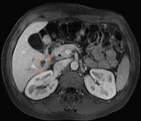
MRI for liver evaluation — hepatocellular carcinoma HCC LI-RADS gadoxetate MRCP liver lesion characterisation
MRI for liver evaluation — with liver-specific gadoxetate contrast (Primovist/Eovist) providing arterial, portal venous, and hepatobiliary phase imaging for HCC diagnosis, LI-RADS characterisation, and MRCP biliary assessment.

Liver MRI for Focal Lesion Characterisation

The primary clinical indication for liver MRI is characterisation of focal liver lesions that are indeterminate on ultrasound or CT. Liver MRI provides superior soft tissue contrast and access to multiple complementary imaging sequences that together allow definitive characterisation of most liver lesions without biopsy. Hepatocellular carcinoma (HCC) is diagnosed on MRI by the LI-RADS (Liver Imaging Reporting and Data System) classification, which categorises liver observations from LR-1 (definitely benign) to LR-5 (definitely HCC) based on imaging features including: arterial phase hyperenhancement (APHE — the lesion enhances more than surrounding liver in the arterial phase, reflecting HCC’s arterial blood supply); washout appearance (the lesion becomes darker than surrounding liver in the portal venous or delayed phase, reflecting washout of contrast); enhancing capsule (a visible rim of enhancement around the lesion, reflecting the fibrous capsule of HCC); and size above 10 mm. An LR-5 observation on MRI with liver-specific contrast (gadoxetate) in a cirrhotic patient is diagnostic for HCC, enabling treatment planning without tissue biopsy. Liver metastases are detected with high sensitivity on DWI sequences (appearing as bright foci) and characterised by their enhancement pattern — colorectal metastases are hypovascular; neuroendocrine metastases are hypervascular. MRI is more sensitive than CT for detecting small hepatic metastases (less than 1 cm), making it the preferred modality for pre-operative metastasis staging before liver surgery. Benign liver lesions — haemangiomas (characteristic T2 bright signal and progressive peripheral enhancement), simple cysts (uniform T2 bright, no enhancement), focal nodular hyperplasia (FNH — T1 isointense with central scar, early enhancement, retention of gadoxetate in the hepatobiliary phase), and hepatic adenoma (fat signal on chemical shift imaging, variable enhancement) — are characterised on MRI by their specific combination of T1, T2, diffusion, and enhancement features. For indeterminate liver lesions found on ultrasound or CT, MRI resolves the diagnostic uncertainty in the majority of cases, directing appropriate management — surveillance, biopsy, or treatment. The blood test context for liver lesion assessment — including AFP as an HCC marker alongside the liver function tests covered in the article on liver function tests explained — provides the biochemical picture alongside MRI findings.
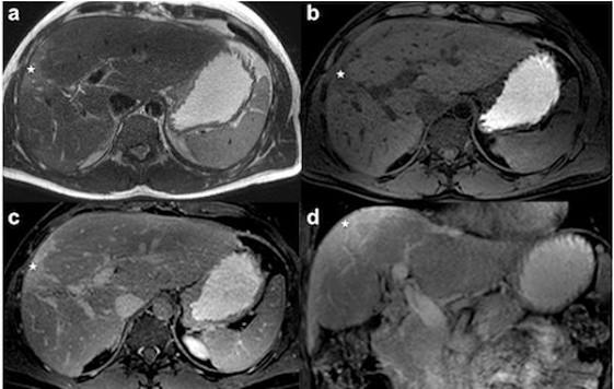
mri-for-liver-evaluation-body — MRCP magnetic resonance cholangiopancreatography bile duct gallstone biliary
MRCP (magnetic resonance cholangiopancreatography) provides non-invasive three-dimensional imaging of the bile ducts and pancreatic duct — detecting stones, strictures, and biliary anatomy without radiation or endoscopy.

Liver-Specific MRI Contrast — Gadoxetate (Primovist/Eovist)

Gadoxetate (sold as Primovist in Europe/Asia and Eovist in North America) is a liver-specific MRI contrast agent that provides both standard dynamic vascular phases (arterial, portal venous) and a unique hepatobiliary phase (HBP) acquired twenty minutes after injection, when functioning hepatocytes take up the contrast agent. This hepatobiliary phase provides functional information about hepatocyte uptake capacity that is unavailable with standard extracellular gadolinium-based contrast: Normal hepatocytes take up gadoxetate, so normal liver parenchyma appears bright on the HBP. HCC — which consists of abnormal tumour cells with reduced hepatocyte organic anion transporter (OATP) expression — does not take up gadoxetate and appears dark (non-enhancing) on the HBP, creating the “washout” appearance that is one of the LI-RADS diagnostic criteria. FNH — which contains functioning hepatocytes — takes up gadoxetate and appears iso- or hyperintense on the HBP, an important distinguishing feature from hepatic adenoma (which does not enhance on HBP). Cholangiocarcinoma — a biliary tumour — shows peripheral enhancement and delayed central fill-in; it does not take up gadoxetate on the HBP. The hepatobiliary phase also provides visualisation of the biliary anatomy, allowing assessment of bile duct patency and biliary communications. Gadoxetate MRI is the most comprehensive and diagnostically accurate liver imaging examination available, combining dynamic vascular characterisation with functional hepatocyte assessment and biliary information in a single investigation. Its use is particularly important in: pre-operative planning before liver resection or transplantation (the HBP provides functional mapping of which liver segments retain function, guiding the extent of safe resection); indeterminate liver lesions in cirrhosis (LI-RADS 3 observations that require follow-up characterisation); and post-treatment response assessment after locoregional therapy (ablation, TACE). The connection between hepatic functional capacity — reflected by gadoxetate HBP uptake — and the synthetic liver blood tests covered in the article on albumin and liver function provides the integrated picture of hepatic reserve assessment.

MRCP — MRI of the Biliary System

Magnetic resonance cholangiopancreatography (MRCP) is a non-invasive MRI technique that produces heavily T2-weighted images in which fluid-containing structures — bile ducts and the pancreatic duct — appear bright, creating a three-dimensional roadmap of the biliary tree and pancreatic ductal anatomy. MRCP has replaced diagnostic ERCP (endoscopic retrograde cholangiopancreatography) for the majority of biliary diagnostic indications because it provides equivalent anatomical information without the risks of ERCP (acute pancreatitis in approximately three to five percent, bile duct perforation, cholangitis). ERCP is now reserved for therapeutic indications — stone extraction, biliary stenting, tissue sampling — where endoscopic access is required for treatment in addition to diagnosis. Clinical indications for MRCP include: suspected common bile duct stones (choledocholithiasis) causing biliary obstruction (jaundice, right upper quadrant pain, elevated bilirubin and ALP); evaluation of biliary strictures (distinguishing benign from malignant strictures, characterising the level of obstruction); primary sclerosing cholangitis (PSC — MRCP characterises the multifocal biliary stricturing pattern of this autoimmune bile duct disease, monitors for dominant stricture development and cholangiocarcinoma); pancreatic ductal abnormalities including IPMN (intraductal papillary mucinous neoplasm — an increasingly detected cystic pancreatic lesion requiring surveillance for malignant transformation); and pre-operative biliary anatomy mapping before liver or pancreatic surgery. The relationship between biliary obstruction and the specific blood test pattern it produces — elevated bilirubin and ALP covered in the articles on bilirubin testing and alkaline phosphatase — provides the clinical trigger for ordering MRCP in a jaundiced patient.

MR Elastography — Measuring Liver Stiffness by MRI

MR elastography (MRE) is a specialised MRI technique that measures liver stiffness — the biomechanical property that increases as fibrosis accumulates and replaces normal soft hepatocytes with rigid scar tissue. MRE uses an acoustic driver (a passive vibrating pad placed on the abdomen) to generate low-frequency mechanical waves in the liver tissue; these waves are detected by a specialised MRI sequence, and wave propagation speed (which correlates with tissue stiffness) is mapped across the entire liver, producing a colour-coded stiffness map (elastogram). Normal liver stiffness on MRE is below 2.5–2.9 kPa depending on the platform and protocol; values above 4–5 kPa indicate significant fibrosis (F2–F3); values above 5–6 kPa indicate cirrhosis (F4). MRE has important advantages over ultrasound-based elastography (FibroScan): it is not affected by body habitus or obesity (the primary limitation of FibroScan), provides a whole-liver stiffness map rather than a single-core measurement, and can detect focal areas of higher stiffness within a heterogeneous cirrhotic liver that might contain an occult tumour. MRE is the most accurate non-invasive tool for fibrosis staging, particularly in obese patients and those with severe ascites where FibroScan is technically unreliable. Its limitations are cost, scan duration, and limited availability compared with FibroScan — making it a second-line tool when FibroScan is technically inadequate rather than a first-line investigation. The relationship between MRE stiffness values, FibroScan results, and histological fibrosis stage connects these non-invasive tools to the biopsy-based fibrosis grading that remains the gold standard, explored further in the context of the liver function tests covered across this series.

Frequently Asked Questions About Liver MRI

What should I expect during a liver MRI?
A liver MRI typically takes thirty to sixty minutes depending on the sequences used and whether contrast is administered. Patients lie still on the MRI table, which slides into the bore of the magnet. The scan involves multiple acquisitions with periods of breath-holding (typically fifteen to twenty-five seconds per acquisition) — the sonographer instructs patients when to hold their breath and when to breathe normally via intercom. The magnet produces loud repetitive knocking and clicking sounds throughout the scan — patients are provided with ear protection. Patients with claustrophobia should inform the radiography team in advance; open MRI systems and short-bore magnets are available in many centres for patients with significant anxiety, and anxiolytic medication can be arranged. Patients with metallic implants — cardiac pacemakers, cochlear implants, certain neurosurgical clips, and some joint replacements — require safety screening before MRI; many modern implants are MR-conditional (safe under specific field strengths and orientations). Intravenous gadolinium-based contrast (for dynamic and hepatobiliary phase imaging) is administered through an IV cannula during the scan. Gadolinium contrast is generally well tolerated; severe allergic reactions occur in approximately 0.01–0.1%, much lower than with iodinated CT contrast. In patients with severe renal impairment (eGFR below 30), gadolinium carries a risk of nephrogenic systemic fibrosis with certain older linear gadolinium agents — modern macrocyclic agents are significantly safer but still require caution in severe renal impairment. No fasting is typically required for liver MRI, though some centres request a four-hour fast to reduce bowel peristalsis artefacts. The hepatobiliary phase MRI using gadoxetate requires a specific timing protocol that the radiology team manages.

How is liver MRI different from a CT scan of the liver?
The key differences between liver MRI and liver CT are: radiation (MRI uses none; CT uses ionising radiation); soft tissue contrast (MRI provides superior soft tissue differentiation and can generate multiple complementary signal contrasts; CT provides one fundamental contrast type supplemented by iodine enhancement); biliary imaging (MRCP from MRI produces a non-invasive cholangiogram; CT cannot replicate this without endoscopic or percutaneous contrast injection); functional hepatocyte assessment (gadoxetate MRI can assess hepatocellular uptake function; CT cannot); and availability and speed (CT is universally available and takes approximately ten seconds of scan time; MRI requires specialist centres and thirty to sixty minutes). The choice between them is guided by the clinical question: CT is preferred for emergency indications, comprehensive staging, and when speed and availability are priorities; MRI is preferred for best possible lesion characterisation, biliary anatomy, functional liver assessment, and radiation-sensitive patients. The clinical picture from blood tests — the liver enzyme patterns covered in the articles on ALT and AST and GGT — always provides the biochemical foundation alongside imaging findings in liver disease assessment.

Sources: ACR LI-RADS — Liver Imaging Reporting · EASL — HCC Clinical Practice Guidelines · RadiologyInfo.org — Liver MRI

MRI for Rectal Cancer and Pelvic Pathology

While the focus of this article is liver evaluation, MRI of the liver is often combined with pelvic MRI in the context of staging colorectal cancer — since rectal cancer staging by MRI pelvis is one of the most important applications of abdominal MRI for digestive disease. MRI pelvis for rectal cancer assessment uses high-resolution T2-weighted sequences to precisely define the relationship between the tumour and the mesorectal fascia (the circumferential resection margin, CRM). CRM involvement — tumour within 1 mm of the mesorectal fascia — predicts a higher risk of local recurrence and is the key determinant of whether pre-operative long-course chemoradiotherapy or short-course radiotherapy is needed before surgery. MRI also assesses tumour T-stage (depth of penetration through the rectal wall), extramural vascular invasion (EMVI — tumour cells in perirectal blood vessels, a prognostic marker associated with systemic metastasis risk), and lymph node involvement in the mesorectal and lateral pelvic compartments. The MRI staging directly determines whether the patient proceeds to primary surgery, neoadjuvant long-course chemoradiation, or short-course radiotherapy — making it one of the most management-directing imaging investigations in colorectal cancer care. Post-treatment MRI restaging after neoadjuvant therapy assesses for pathological complete response (pCR), which may allow non-operative management (watch-and-wait) as an alternative to surgery in carefully selected patients. The connection between rectal cancer investigation and the broader gastrointestinal diagnostic framework — including the stool tests that may have prompted investigation — is covered in the article on stool tests for digestive health.

When Liver MRI Cannot Be Performed

Liver MRI, despite its diagnostic superiority for many indications, cannot be performed in a subset of patients due to absolute or relative contraindications. Absolute contraindications include: cochlear implants (most are MR-unsafe); certain cardiac pacemakers and implantable cardioverter-defibrillators (older non-MR-conditional devices — many modern devices are now MR-conditional); certain intracranial aneurysm clips; and intraorbital metallic foreign bodies (occupational exposure in metalworkers requires skull X-ray screening). Relative contraindications requiring safety assessment include: knee or hip prostheses (most modern orthopaedic implants are MR-safe or MR-conditional); breast tissue expanders and implants (device-specific assessment required); neurostimulators; insulin pumps; and retained bullet or shrapnel fragments. For patients with contraindications to MRI, CT with multi-phase liver protocol and contrast-enhanced ultrasound (CEUS) provide the primary alternative characterisation pathways. CEUS — ultrasound combined with intravenous microbubble contrast — provides real-time vascular characterisation of liver lesions comparable to CT and MRI for many indications, with the additional advantages of no radiation and real-time guidance capability. Claustrophobia is not an absolute contraindication — open MRI systems with wider bores are available in many centres, anxiolytic premedication is routinely used for patients with significant anxiety, and general anaesthesia is available for those who cannot tolerate the scan awake. The clinical decision of which imaging modality to use when MRI is not possible is made by the requesting clinician in discussion with the radiology team — the goal is always to select the modality that best answers the clinical question for that specific patient. Understanding the full investigative picture — from blood tests through ultrasound to MRI — is covered across the liver and digestive health series, with the overview article on abdominal ultrasound providing the foundation imaging context.

MRI Findings and Their Clinical Significance

MRI findings in the liver are reported using standardised systems and terminology designed to ensure consistent communication between radiologists and clinicians. For liver lesion characterisation, the LI-RADS system (described above) provides a standardised vocabulary for lesion reporting in cirrhotic and at-risk patients. For general liver assessment, the MRI report will describe: Liver size and morphology — as on CT, liver enlargement or cirrhotic atrophy/hypertrophy patterns; Parenchymal signal — T1 and T2 signal characteristics that reflect fat content, iron deposition (haemochromatosis produces low T2 signal in a characteristic pattern — affecting both the liver and spleen in secondary haemosiderosis, or predominantly liver in HFE-related hereditary haemochromatosis), fibrosis, and inflammatory change; Focal lesions — characterised by their T1 and T2 signal, DWI restriction, and enhancement pattern as described; Biliary system — MRCP findings of ductal calibre, filling defects (stones appear as dark foci against bright bile on MRCP), strictures, and anatomical variants; and Adjacent organs — the spleen, pancreas, kidneys, adrenal glands, and vascular structures are assessed as part of the comprehensive MRI examination. For patients receiving their first liver MRI report, understanding that descriptive MRI terminology (e.g., “T2 hyperintense lesion with restricted diffusion and arterial phase hyperenhancement”) describes the imaging characteristics rather than making a direct pathological diagnosis helps bridge the gap between the technical report and its clinical meaning. The treating clinician or a radiologist will translate these imaging descriptors into clinical conclusions and management recommendations. The biochemical context from liver blood tests — including the viral hepatitis markers covered in hepatitis blood tests explained and the synthetic function markers in albumin and liver function — provides the essential clinical context for interpreting MRI findings accurately.

Liver MRI represents the current apex of non-invasive liver imaging — combining the absence of ionising radiation with the best available soft tissue contrast, functional hepatocyte assessment through liver-specific contrast, comprehensive biliary anatomy via MRCP, and mechanistic fibrosis quantification through MR elastography. For patients with known or suspected liver disease requiring the most accurate possible non-invasive characterisation, MRI provides information that no other imaging modality can replicate. Its appropriate use — as a second-line characterisation tool for indeterminate lesions, as a definitive staging tool for biliary and hepatic malignancy, and as a functional assessment tool for pre-surgical planning — reflects the evidence base that has established liver MRI as the gold standard imaging modality for liver disease assessment in carefully selected clinical indications.

For patients navigating a liver disease diagnosis that involves liver MRI as part of their workup — whether for a newly discovered liver lesion, cirrhosis staging, or pre-surgical assessment — understanding the distinctive capabilities of MRI relative to ultrasound and CT, and how each investigation complements the others in building a complete diagnostic and monitoring picture, enables informed engagement with the clinical pathway. The combination of liver function blood tests covered across this series — including the detailed explanations of ALT and AST, GGT, bilirubin, and the CT and ultrasound imaging articles — provides the integrated clinical framework within which liver MRI findings take on their full diagnostic and prognostic significance. The technical complexity of MRI is matched by the depth of clinical information it provides; for the right clinical question, it remains the most powerful non-invasive tool available for liver disease assessment.

Patients and their families who want to understand the complete arc of liver disease investigation — from the initial blood tests that first raise concern, through the ultrasound that characterises the liver, to the CT and MRI that define the pathology — will find that each investigation builds on the last. The liver ultrasound article and the CT scan article in this series provide the contextual framework into which liver MRI results fit, ensuring the full picture is accessible without requiring specialist radiology training.

3 thoughts on “MRI for Liver Evaluation

  1. Daniel Tran says:

    Really helpful overview. My hepatologist ordered a liver MRI after my AFP came back mildly elevated and I had no idea what to expect. The section on LI-RADS categories makes the radiology report much less intimidating — I finally understand what LR-3 means versus LR-4.

    • Horizon Health Guide says:

      Thank you Daniel — that’s exactly the situation where the LI-RADS section is most useful. An LR-3 finding represents an intermediate probability of HCC and typically leads to a follow-up MRI at 3–6 months, while LR-4 carries a higher probability and may prompt biopsy or MDT discussion depending on clinical context. If your hepatologist has recommended follow-up imaging, the interval and modality they’ve chosen reflects that LI-RADS category alongside your broader clinical picture. Wishing you all the best with your follow-up.

  2. Margaret Osei says:

    I was nervous about the MRI because I have a hip replacement, but my radiographer confirmed the implant is MR-conditional and the scan went ahead fine. The section on contraindications was reassuring to read beforehand. The MRCP images were impressive — you could see exactly where the bile duct stricture was without any invasive procedure.

Leave a Reply

Your email address will not be published. Required fields are marked *