
A CT scan (computed tomography scan, also called a CAT scan) uses X-rays combined with computer processing to create detailed cross-sectional images of the body’s internal structures. In digestive and liver medicine, CT scanning is one of the most important and frequently used diagnostic and monitoring tools — it provides superior spatial resolution compared with ultrasound, can image all abdominal and pelvic organs including bowel (which is largely inaccessible to standard ultrasound), and can be performed with intravenous contrast that enables dynamic characterisation of lesion vascularity for definitive diagnosis of liver tumours, bowel pathology, and vascular disease. Understanding what a CT scan can show for different digestive and liver conditions, how it is performed, what the risks of radiation and contrast agents are, and how it fits into the diagnostic pathway alongside ultrasound and MRI helps patients understand why this investigation was ordered and how to make sense of CT scan reports.
CT scanning works by rotating an X-ray source around the patient while detectors on the opposite side measure the attenuation of the X-ray beam as it passes through different tissues. Bone attenuates X-rays strongly (appears bright white), air attenuates poorly (appears black), and soft tissues — including the liver, bowel wall, pancreas, and lymph nodes — attenuate at intermediate levels (appearing in various shades of grey). The data from multiple rotation angles is reconstructed by computer into a three-dimensional dataset from which cross-sectional images in any plane can be generated. Modern multi-detector CT scanners can image the entire abdomen and pelvis in a single breath-hold of approximately ten seconds, providing isotropic (equal resolution in all directions) images that can be reformatted in any plane without loss of resolution. This capability makes CT the modality of choice for comprehensive abdominal assessment, staging of malignancy, evaluation of acute abdominal emergencies, and characterisation of focal liver lesions that are indeterminate on ultrasound.
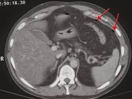
CT Scan for Liver Disease and Focal Liver Lesions
The liver is one of the primary targets of abdominal CT scanning, and contrast-enhanced CT with multi-phase liver protocol is the standard modality for characterising indeterminate liver lesions found on ultrasound and for staging liver malignancy. A multi-phase liver CT includes: pre-contrast phase (assessing baseline density and identifying calcification or haemorrhage); arterial phase (twenty-five to thirty-five seconds after contrast injection — captures early arterial enhancement in hypervascular lesions); portal venous phase (sixty to seventy seconds — the primary phase for liver parenchymal assessment and most lesion detection); and sometimes a delayed phase (three to five minutes — helps characterise fibrous or sclerotic tumours). Hepatocellular carcinoma (HCC) has a characteristic CT appearance called “arterial enhancement with washout” — it appears bright in the arterial phase (hyperenhancing due to its arterial blood supply) and darker than the surrounding liver in the portal venous or delayed phase (washout appearance). This specific enhancement pattern is diagnostic for HCC on CT in a cirrhotic liver, enabling non-invasive diagnosis without biopsy according to both EASL and AASLD guidelines. Liver metastases from colorectal cancer typically appear as hypovascular lesions — darker than liver parenchyma in the portal venous phase — while neuroendocrine tumour metastases are hypervascular and best seen in the arterial phase. Haemangiomas show characteristic peripheral nodular enhancement with progressive centripetal fill-in — a pattern seen on delayed phase imaging. CT is also used to stage known liver malignancy — assessing the size and number of liver lesions, involvement of hepatic veins or portal vein (vascular invasion), extrahepatic disease (lymph nodes, peritoneal deposits, lung metastases), and assessing feasibility of surgical resection by calculating future remnant liver volume. The blood test markers of liver function — covered in the articles on liver function tests and albumin testing — provide the biochemical baseline alongside CT staging findings.
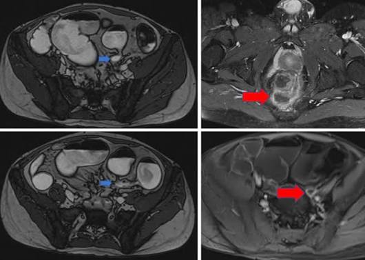
CT Scan for Pancreatic and Biliary Disease
The pancreas — largely inaccessible to ultrasound due to overlying bowel gas — is optimally assessed by CT, making abdominal CT with dedicated pancreatic protocol the investigation of choice when pancreatic pathology is suspected. A pancreatic protocol CT uses specific timing to maximise enhancement of the pancreatic parenchyma (pancreatic phase, approximately forty to fifty seconds after contrast) alongside the arterial and portal venous phases, providing optimal lesion-to-parenchyma contrast for detecting pancreatic masses. Pancreatic ductal adenocarcinoma (PDAC) — the most clinically important pancreatic tumour — typically appears as a hypovascular (darker than surrounding pancreas) mass in the head, body, or tail of the pancreas, often with upstream pancreatic duct dilation. The critical CT assessment for pancreatic cancer is vascular involvement — whether the superior mesenteric artery, coeliac axis, portal vein, or superior mesenteric vein is encased or abutted by tumour, since vascular involvement determines surgical resectability. Resectable pancreatic cancer (no vascular involvement) has the best prognosis with surgery; borderline resectable and unresectable tumours require oncological discussion for neoadjuvant therapy or palliation. Acute pancreatitis is assessed by CT when clinical diagnosis is uncertain, when the patient is severely unwell, or when complications are suspected. The CT Severity Index (CTSI) grades pancreatitis severity from the extent of pancreatic inflammation and necrosis — the presence of pancreatic necrosis (non-enhancing pancreatic parenchyma) is the most important prognostic marker, associated with significantly higher morbidity and mortality. Chronic pancreatitis produces characteristic CT findings including pancreatic parenchymal calcification (pathognomonic), pancreatic duct dilation and irregularity, and pancreatic atrophy. The stool elastase test covered in the article on stool tests for digestive health provides the functional counterpart to CT’s structural assessment of chronic pancreatitis. Biliary disease assessment by CT includes detection of biliary dilation (the level and cause of obstructive jaundice), cholangiocarcinoma staging, and post-operative biliary complication evaluation. However, MRCP (MR cholangiopancreatography) is superior to CT for visualising the biliary tree and detecting common bile duct stones, and is typically preferred for biliary pathway evaluation when the jaundice aetiology is uncertain.
CT for Colorectal Cancer and Bowel Disease
CT plays a critical role in both colorectal cancer staging and bowel disease evaluation. CT colonography (virtual colonoscopy, CTC) uses a thin rectal tube to insufflate the colon with CO₂ gas, followed by a low-dose CT scan of the distended colon. Computer software creates a three-dimensional fly-through of the colon lumen, allowing detection of colorectal polyps (above 6 mm sensitivity approximately ninety percent) and colorectal cancer without conventional colonoscopy. CT colonography is used for colorectal cancer screening in patients unsuitable for conventional colonoscopy (high anaesthetic risk, anticoagulation, incomplete prior colonoscopy), has no sedation requirement, and simultaneously evaluates extracolonic abdominal organs for incidental pathology. Unlike conventional colonoscopy, it cannot remove polyps or take biopsies — a positive CTC finding (polyp above 10 mm) requires follow-up conventional colonoscopy for polypectomy. CT colonography is included in some national colorectal cancer screening pathways as an alternative to colonoscopy. CT for colorectal cancer staging follows the TNM staging system: T (tumour depth through the bowel wall) is better assessed by MRI pelvis for rectal cancer; N (lymph node involvement) and M (distant metastases — most commonly liver and lung) are assessed by CT chest, abdomen, and pelvis. Staging CT is essential before any colorectal cancer surgery to guide the extent of resection and the need for neoadjuvant (pre-operative) chemotherapy or radiotherapy. CT for acute bowel conditions — including bowel obstruction, intestinal ischaemia, diverticulitis, appendicitis, and perforated viscus — is the primary imaging modality in emergency presentations with acute abdominal pain. CT can identify the cause, site, and complications of acute bowel disease within minutes, guiding whether the patient needs emergency surgery, conservative management with antibiotics, or interventional radiology drainage of an abscess. The symptom of altered bowel habit and bleeding that prompts colorectal investigation is covered in the article on when digestive symptoms need medical attention.
Radiation, Contrast, and CT Scan Safety
CT scanning uses ionising radiation — the dose from a typical abdominal and pelvic CT is approximately 8–14 mSv, equivalent to approximately three to five years of natural background radiation. The risk of radiation-induced cancer from a single abdominal CT is very low in absolute terms (estimated at approximately 1 in 2000 to 1 in 5000 for adults) and must always be weighed against the clinical benefit of the diagnostic information obtained. For patients with potentially serious conditions — suspected malignancy, acute abdominal emergency, biliary obstruction — the benefit of accurate CT diagnosis vastly outweighs the small radiation risk. For younger patients and for conditions where repeated CT imaging may be needed, radiation dose minimisation using low-dose protocols and consideration of alternative modalities (MRI, ultrasound) where clinically appropriate is good practice. Intravenous iodinated contrast — used in most diagnostic abdominal CT scans — carries a small risk of allergic reaction (mild reactions in approximately 1–3%, severe anaphylaxis in approximately 0.02–0.04%) and contrast-induced nephropathy (kidney function impairment, more significant in patients with pre-existing chronic kidney disease — eGFR below 30 requires careful risk-benefit assessment and pre-hydration protocols). Patients should inform the radiology team of prior contrast reactions, renal impairment, metformin use (held twenty-four to forty-eight hours around contrast administration), thyroid disease, and pregnancy. Patients who are pregnant should avoid CT whenever possible due to foetal radiation risk — ultrasound or MRI are the preferred alternatives in pregnancy. The diagnostic context in which CT is ordered — including the biochemical markers covered in the articles on ALT and AST, bilirubin, and GGT — provides the integrated assessment that determines whether CT is the appropriate imaging choice.
Frequently Asked Questions About CT Scans for Digestive Health
Do I need to prepare for an abdominal CT scan?
Preparation requirements for CT vary by indication. For most abdominal CT scans with intravenous contrast, patients are asked to fast for four to six hours before the scan to reduce the risk of aspiration if contrast causes nausea. Oral contrast (a dilute contrast solution drunk before the scan to opacify the bowel) may be given in some CT protocols to distinguish bowel loops from other abdominal structures. For CT colonography, full bowel preparation (similar to colonoscopy prep, using laxatives the day before to clear the colon of faecal residue) is required to allow adequate assessment of the colon wall and to prevent stool polyp mimics. Some departments use faecal tagging (drinking dilute contrast in the days before the scan so stool is contrast-labelled) as an alternative to full preparation. For CT without contrast — sometimes used for kidney stone detection or acute abdominal pain assessment — no fasting or preparation is typically required. Patients should arrive with their referral information, a list of current medications, and any prior imaging for comparison.
My CT showed an incidental finding — should I be worried?
Incidental findings — abnormalities found on CT that were not the primary reason for the scan — are extremely common, identified in approximately thirty to forty percent of all abdominal CT scans. The vast majority are benign: simple liver cysts, small haemangiomas, benign renal cysts, adrenal adenomas, small pulmonary nodules. The challenge is that some incidental findings require follow-up imaging to confirm their benign nature or to detect any change that would indicate a more significant lesion. Standardised reporting guidelines (such as the ACR Incidental Findings Committee white papers) provide radiologists with evidence-based recommendations for when incidental findings require follow-up, when they can be ignored, and what modality is most appropriate. When you receive a CT report with an incidental finding, your clinician will explain what was found, its likely nature, and what if any follow-up is recommended. An incidental liver lesion below 1 cm in a low-risk patient with a normal liver and no known malignancy history can usually be followed up with ultrasound at six to twelve months; the same lesion in a patient with known malignancy requires urgent MRI characterisation. The clinical context provided by blood tests — including the hepatitis markers in the hepatitis blood tests article and the H. pylori testing covered in the H. pylori test guide — provides the clinical framework within which CT findings are interpreted.
Sources: ACR Appropriateness Criteria — Abdominal CT Imaging · RadiologyInfo.org — Abdominal CT · NIDDK — Diagnostic Tests Overview
CT vs MRI vs Ultrasound: Choosing the Right Imaging Modality
The choice between CT, MRI, and ultrasound for digestive and liver imaging depends on multiple factors that reflect the specific clinical question, patient factors, and available resources. Understanding how these modalities compare helps patients understand why one was chosen over another for their particular situation. Ultrasound is first-line for suspected gallstones, initial liver assessment, suspected biliary obstruction (bile duct dilation), evaluation of right upper quadrant pain, and as a screening tool for liver disease and HCC surveillance. It is the safest modality (no radiation), most widely available, and least expensive. Its limitations include operator dependence, bowel gas interference, and limited resolution for deep structures. CT is preferred over ultrasound when: comprehensive abdominal assessment including bowel is required (staging, acute emergency); retroperitoneal pathology is suspected (pancreatic mass, lymphadenopathy); lesions found on ultrasound need contrast characterisation; and whole-body staging for known or suspected malignancy is needed. CT is faster than MRI and available in all hospitals — essential for emergency indications. Its limitations are radiation exposure and iodine contrast risks. MRI is preferred over CT when: the best possible soft tissue characterisation is needed (liver lesion characterisation, biliary anatomy with MRCP, rectal cancer T-staging); radiation avoidance is a priority (younger patients, pregnant women, repeat imaging); and functional imaging of liver stiffness (MR elastography) is required. MRI is slower (twenty to forty-five minutes), more expensive, not available everywhere, and not suitable for patients with certain metallic implants (pacemakers, cochlear implants, some aneurysm clips). The three modalities are therefore complementary rather than competing — each occupies a specific position in the investigation pathway determined by the clinical question and patient factors. For digestive and liver symptoms that trigger imaging, the clinical markers covered in the articles on fatigue and liver health and dark urine and liver health provide the clinical context that determines which modality is most appropriate.
CT in the Staging of Gastrointestinal Malignancy
CT staging of gastrointestinal cancers is one of the most important clinical applications of abdominal CT — it determines treatment planning, guides surgical decision-making, and provides the baseline against which treatment response is assessed. For oesophageal and gastric cancer, CT chest, abdomen, and pelvis assesses tumour extent, regional lymph node involvement, and distant metastases (liver being the most common site). CT is supplemented by PET-CT in many centres to detect occult distant metastases that may alter treatment intent from curative to palliative. For colorectal cancer, CT chest, abdomen, and pelvis is the primary staging tool for colon cancer (T, N, M staging), with MRI pelvis used specifically for rectal cancer to determine the tumour relationship to the mesorectal fascia (circumferential resection margin — a key determinant of whether pre-operative chemoradiotherapy is needed). CT liver assessment in colorectal cancer is critical: up to forty percent of colorectal cancer patients develop liver metastases, and CT-identified liver metastases determine whether the patient is resectable (potentially curative liver resection in appropriate candidates), suitable for liver-directed therapy (thermal ablation, SIRT), or requires systemic chemotherapy alone. For hepatocellular carcinoma, CT chest, abdomen, and pelvis with multi-phase liver protocol provides the oncological staging and assesses vascular involvement that determines eligibility for liver transplantation (Milan criteria — one lesion below 5 cm, or up to three lesions none above 3 cm), surgical resection, or locoregional therapy. The biochemical picture alongside CT staging — including the coagulation and synthetic function markers covered in the article on liver function tests — provides the physiological assessment that complements the anatomical staging CT provides.
CT-Guided Procedures and Interventional Applications
Beyond diagnostic imaging, CT is used to guide minimally invasive procedures — CT-guided biopsy and CT-guided drainage are two of the most important. CT-guided liver biopsy allows precise targeting of focal liver lesions that are too deep, too small, or in a technically difficult location for ultrasound-guided biopsy. A biopsy needle is advanced under real-time CT fluoroscopy guidance through the abdominal wall directly into the target lesion, and tissue is obtained for histopathological analysis. This enables diagnosis of liver lesions that cannot be diagnosed by imaging characteristics alone — indeterminate lesions below the threshold for non-invasive HCC diagnosis, suspected metastases where the primary site is uncertain, cholangiocarcinoma, and diffuse liver disease where the biopsy target is the liver parenchyma rather than a focal lesion. CT-guided drainage of abdominal abscesses, infected pancreatic necrosis (necrosectomy), and pleural effusions uses CT to plan and guide percutaneous drain placement — draining infected collections that would otherwise require open surgery. Tumour ablation under CT guidance — including radiofrequency ablation (RFA), microwave ablation (MWA), and cryoablation of liver metastases and small HCC — uses CT to plan the ablation probe trajectory and confirm adequate ablation zone coverage. These procedures expand CT’s role from purely diagnostic to therapeutically interventional, reflecting the full scope of its application in digestive and liver disease management. The diagnostic pathway that leads to these interventions — from initial blood test abnormalities through imaging characterisation to procedural intervention — represents the full arc of liver disease investigation in which CT plays a central and indispensable role. Understanding how individual investigations fit into this pathway — from the initial blood tests covered across the liver and digestive health series to the advanced imaging and procedural interventions — equips patients to navigate their care with confidence and to engage meaningfully with the clinical decisions being made on their behalf.
CT scanning’s combination of speed, comprehensive anatomical coverage, and contrast-based lesion characterisation makes it the backbone of gastrointestinal and liver disease imaging in both elective and emergency settings. For patients receiving an abdominal CT — whether for initial investigation of symptoms, staging of a known diagnosis, follow-up of a prior finding, or emergency assessment — understanding what the investigation provides and how its findings integrate with blood test results and clinical symptoms enables informed participation in the clinical decision-making process. The liver blood tests covered in the series of articles on this site — from ALT and AST to alkaline phosphatase — provide the biochemical foundation on which CT imaging findings are built, giving the complete diagnostic picture that guides management of digestive and liver disease across the full spectrum from early detection to advanced disease monitoring.
For patients navigating a diagnosis that involves multiple imaging investigations over time — a liver mass under surveillance, a bowel cancer being treated, a chronic pancreatic condition being monitored — the accumulation of CT reports alongside blood tests, endoscopy results, and clinical assessments can feel overwhelming. A useful framework is to think of each investigation as answering one specific question: the CT answers the structural anatomical question; the blood tests answer the biochemical functional question; endoscopy answers the mucosal surface question; and biopsy answers the histological tissue question. Together these form a multi-dimensional diagnostic picture that no single investigation can provide alone. Understanding each test’s specific contribution — as explored across this series of liver and digestive health articles — transforms the diagnostic process from a series of opaque procedures into a coherent, comprehensible investigation pathway aimed at accurate diagnosis, precise staging, and optimal treatment selection.
I had an abdominal CT after my ultrasound found an indeterminate liver lesion. The CT showed arterial enhancement with washout in a segment 5 nodule against a background of cirrhosis — my hepatologist explained this is diagnostic for HCC without needing a biopsy. I am now being assessed for liver transplant. The speed from ultrasound finding to CT characterisation to treatment planning was about three weeks — the system worked exactly as described here.
Brian, LI-RADS 5 (arterial enhancement with washout in cirrhosis) is one of the most powerful diagnostic findings in imaging — it genuinely changes the diagnostic certainty to a point where tissue biopsy is no longer required before treatment planning. The efficiency you describe — ultrasound detection to CT characterisation to transplant assessment in three weeks — represents the surveillance programme working at its best. Your story will encourage others to maintain their six-monthly surveillance appointments.
The pancreatic cancer section is really important. My father presented with jaundice and weight loss — CT showed a 3cm mass in the pancreatic head with involvement of the superior mesenteric vein. That involvement meant it was borderline resectable, not immediately operable. He had chemotherapy first to try to downstage the tumour before surgery. I never understood what ‘borderline resectable’ meant until reading this. Thank you for explaining the vascular assessment so clearly.