
Liver biopsy is a procedure in which a small sample of liver tissue — typically a core approximately 2–3 cm long and 1–2 mm in diameter — is obtained for histopathological examination. It remains the most information-rich single investigation in hepatology: a well-sampled liver biopsy can simultaneously assess the stage of fibrosis (degree of scarring), grade of inflammation (necroinflammatory activity), degree of steatosis (fat content), the presence of specific histological features that identify the underlying aetiology (viral hepatitis, autoimmune hepatitis, metabolic steatohepatitis, primary biliary cholangitis, primary sclerosing cholangitis, haemochromatosis, Wilson’s disease), and the presence of complications such as dysplastic nodules or early hepatocellular carcinoma. For patients who have been told they need a liver biopsy — or who are considering whether to accept one after non-invasive tests have been inconclusive — understanding the clinical purpose of the biopsy, what the procedure involves, the information it provides and what the results mean, and the realistic risk profile is essential to informed decision-making.
Liver biopsy is no longer the routine first-line investigation for liver disease assessment — the development and validation of non-invasive fibrosis markers (FibroScan, serum-based scores, and MR elastography as described in the FibroScan and MRI articles in this series) has allowed biopsy to be reserved for situations where non-invasive assessment is inadequate, discordant, or where specific histological information is required that non-invasive testing cannot provide. When liver biopsy is recommended, it is because the clinical question cannot be fully answered by other means — and the information obtained directly shapes diagnosis and treatment decisions in ways that blood tests and imaging cannot replicate.
When Liver Biopsy Is Indicated
Liver biopsy is recommended in specific clinical situations where the histological information cannot be obtained by alternative means and is necessary for clinical management. The key indications in current practice are:
Discordant non-invasive fibrosis assessment: When FibroScan, serum fibrosis markers (FIB-4 index, APRI score), and clinical assessment give conflicting results — for example, a low FIB-4 score but a liver stiffness suggesting advanced fibrosis — biopsy resolves the discordance by providing direct histological fibrosis staging. Discordance is particularly common in patients with confounders that elevate liver stiffness independently of fibrosis (acute hepatitis, biliary obstruction, right heart failure, active alcohol use), in whom non-invasive results may overestimate fibrosis stage.
Diagnosis of autoimmune liver disease: Autoimmune hepatitis (AIH) requires histological confirmation for diagnosis — the simplified International Autoimmune Hepatitis Group (IAIHG) scoring system incorporates histological features (interface hepatitis, rosette formation, plasma cell infiltrate) that are essential for diagnostic certainty. AIH treatment with immunosuppression (prednisolone and azathioprine) is guided by histological disease activity; assessment of complete histological remission before treatment withdrawal requires biopsy in most guidelines. Primary biliary cholangitis (PBC) and primary sclerosing cholangitis (PSC) — though diagnosed primarily on biochemistry and cholangiography — may require biopsy for staging or when serological and imaging findings are atypical. The liver function test pattern in PBC and PSC — cholestatic pattern with elevated ALP and GGT — is covered in the series articles on GGT testing and liver function tests.
Investigation of suspected overlap syndromes: When clinical features suggest more than one liver disease simultaneously — for example, autoimmune hepatitis overlapping with PBC, or MASLD occurring in a patient with chronic hepatitis B — biopsy is the only investigation that can assess the contributions of each disease process to the overall histological picture. Management of overlap syndromes depends on which disease is predominant histologically.
Unexplained liver abnormalities: Persistently elevated liver enzymes (ALT, AST, ALP, GGT) or imaging abnormalities that remain unexplained after a comprehensive non-invasive assessment — including viral hepatitis serology, autoimmune liver markers, metabolic workup, and appropriate imaging — may be investigated with biopsy when the clinical concern for significant liver disease is sufficient and the non-invasive workup is negative or inconclusive.
Assessment of drug-induced liver injury (DILI): When a specific hepatotoxic drug is the suspected cause of liver disease but cannot be confirmed by clinical criteria alone, biopsy can characterise the histological pattern of injury (hepatocellular, cholestatic, or mixed; zonal necrosis pattern suggesting ischaemia or specific hepatotoxins; granulomatous hepatitis associated with certain drug classes) and help differentiate DILI from coincidental underlying liver disease. Liver tumour assessment: Liver masses that are indeterminate on CT and MRI and that do not fit standard imaging criteria for HCC or haemangioma may require biopsy for histological characterisation — particularly metastatic disease, cholangiocarcinoma, hepatic lymphoma, and rare benign lesions. This decision involves careful consideration of biopsy risk versus diagnostic certainty, particularly for potentially resectable lesions where needle tract seeding is a theoretical concern (though practically rare).
How Liver Biopsy Is Performed
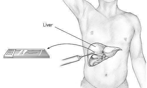
The most common technique is percutaneous liver biopsy under ultrasound guidance. The patient lies supine or in the left lateral position; the right lobe of the liver is identified by ultrasound, and the biopsy site — typically the right upper quadrant in the mid-axillary line — is selected to avoid major bile ducts and blood vessels. The skin is cleaned and local anaesthetic (lidocaine) is infiltrated into the skin, subcutaneous tissue, and liver capsule. A biopsy needle — most commonly a semi-automated Menghini-type aspiration needle or a spring-activated Tru-Cut (core biopsy) needle, typically 16 or 18 gauge — is advanced to the liver under real-time ultrasound guidance, and the core of tissue is obtained in a single pass lasting less than one second. The core biopsy approach is preferred in most centres because it provides better tissue architecture preservation for histological assessment, particularly for fibrosis staging. The procedure typically takes fifteen to twenty minutes including preparation, and patients undergo a period of observation afterwards.
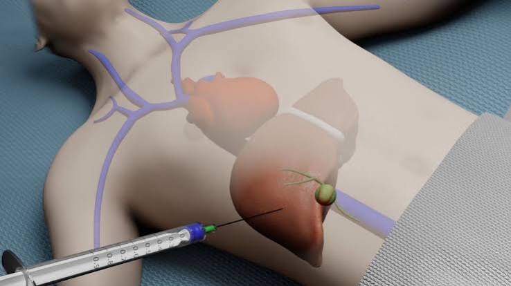
Alternatives to percutaneous biopsy: Transjugular liver biopsy (TJLB) is performed when percutaneous biopsy is contraindicated due to coagulopathy, thrombocytopaenia, or ascites that make the percutaneous approach unsafe. A catheter is inserted via the internal jugular vein, advanced through the right heart into the hepatic veins, and a biopsy needle is passed through the hepatic vein wall into the liver parenchyma — the biopsy is taken from within the hepatic vein, so any post-biopsy bleeding enters the venous circulation rather than the peritoneal cavity. This approach allows biopsy in patients with severe coagulopathy (INR above 1.5) or significant thrombocytopaenia (platelets below 50) who cannot receive adequate pre-procedure correction. Transjugular biopsy also provides simultaneous measurement of the hepatic venous pressure gradient (HVPG) — a direct measure of portal hypertension that is the gold standard for assessing cirrhosis severity and monitoring portal hypertension treatment response. Laparoscopic liver biopsy is performed under general anaesthesia when direct visualisation of the liver surface is required — in patients with suspected liver surface disease, in cirrhotic patients where the liver surface nodularity needs direct assessment, or when peritoneal sampling is also required. CT-guided biopsy is used for focal liver lesions that are not clearly visible on ultrasound or that require CT targeting for precise needle placement.
Preparing for a Liver Biopsy
Pre-biopsy preparation involves blood test assessment, medication review, and fasting. A clotting screen (INR/prothrombin time) and platelet count are checked before the procedure — most centres require an INR below 1.5 and platelets above 50 × 10⁹/L for percutaneous biopsy; values outside these thresholds trigger consideration of transjugular biopsy or pre-procedure correction (fresh frozen plasma, vitamin K for warfarin reversal, platelet transfusion). Anticoagulants are withheld before biopsy — warfarin for five days (with INR check on the day of procedure), DOACs for forty-eight hours in most cases; antiplatelet agents including aspirin are typically withheld for five to seven days. NSAIDs should be stopped one week before biopsy due to their platelet-inhibiting effects. Patients fast for four to six hours before the procedure. A responsible adult escort is required for discharge, particularly if sedation is used.
Patients are asked about allergies (particularly to local anaesthetic), previous biopsy complications, significant comorbidities that might affect the procedure or recovery, and current medications. Informed consent is obtained covering the procedure, indication, alternatives, and risks. Most percutaneous liver biopsies are performed as day-case procedures — patients attend, undergo the procedure, are observed for four to six hours, and are discharged home the same day if no complications arise. Some centres perform biopsies as inpatient procedures (overnight observation) in patients with higher complication risk.
Understanding Liver Biopsy Results
Liver biopsy results are reported by a histopathologist who examines stained tissue sections under light microscopy. Multiple staining techniques are used: haematoxylin and eosin (H&E) for general tissue architecture and cellular morphology; Masson’s trichrome or Sirius red for fibrosis (collagen stains blue or red against the pink hepatocytes, allowing fibrosis to be visualised and staged); Perls’ Prussian blue for iron (identifies haemosiderosis in haemochromatosis and secondary iron overload); rhodanine for copper (Wilson’s disease); periodic acid-Schiff with diastase (PAS-D) for alpha-1 antitrypsin globules; immunohistochemistry for viral antigens, lymphocyte markers, and tumour markers.
Fibrosis staging is reported using a validated scoring system — the METAVIR system (F0–F4) is most widely used: F0 = no fibrosis; F1 = portal fibrosis without septa; F2 = portal fibrosis with few septa (significant fibrosis); F3 = numerous septa without cirrhosis (advanced fibrosis); F4 = cirrhosis. The Ishak scoring system (0–6) and the Batts-Ludwig system are also used depending on centre preference and disease context. For MASLD, the NAFLD Activity Score (NAS) from Kleiner incorporates steatosis (0–3), lobular inflammation (0–3), and hepatocellular ballooning (0–2) — a NAS of 5 or more is associated with NASH (non-alcoholic steatohepatitis), the more severe form of MASLD. Inflammation grading (necroinflammatory activity) is reported alongside fibrosis stage — high-grade inflammation indicates active ongoing liver injury and may require treatment even in patients with low fibrosis stage. Understanding both the stage and grade — the structural consequence of past injury and the level of current inflammatory activity — provides the complete picture that clinicians need to make treatment decisions. The non-invasive tests that provide surrogate assessments of these biopsy parameters — FibroScan for fibrosis stage, ALT for inflammatory activity — are covered across the relevant articles in this series including the ALT and AST article and the FibroScan guide.
Risks and Complications of Liver Biopsy
Liver biopsy carries a small but real risk of complications that must be weighed against its diagnostic value. The overall serious complication rate for percutaneous biopsy in appropriately selected patients is approximately one in two hundred to one in five hundred. Pain: The most common experience after biopsy — right upper quadrant pain or referred right shoulder pain (from diaphragmatic irritation) — occurs in a majority of patients and is typically mild to moderate, lasting one to four hours and managed with paracetamol or mild opioid analgesia. Haemorrhage: The most serious complication — bleeding into the peritoneal cavity or into the liver parenchyma (intrahepatic haematoma) — occurs in approximately one in two hundred to one in five hundred procedures. The majority of post-biopsy haemorrhages are minor and self-limiting; clinically significant haemorrhage requiring transfusion or intervention occurs in approximately one in one thousand procedures and is fatal in approximately one in ten thousand. Risk factors for haemorrhage include coagulopathy, thrombocytopaenia, cirrhosis, multiple passes, and biopsy of vascular lesions. The four-to-six hour post-procedure observation period exists primarily to monitor for haemorrhage — most significant post-biopsy bleeding presents within this window. Biliary complications: Inadvertent puncture of a bile duct can cause biloma (bile leak into the peritoneum) or haemobilia (bleeding into the biliary system) — both rare but manageable with interventional radiology or endoscopic techniques. Infection: Rare in immunocompetent patients with adequate aseptic technique. Pneumothorax: Very rare; occurs when the needle inadvertently traverses the pleural space. Patients should seek urgent medical review if they develop worsening pain, shortness of breath, dizziness, haemoptysis, or feel unwell after biopsy discharge.
Frequently Asked Questions About Liver Biopsy
Non-invasive tests showed significant fibrosis — do I still need a biopsy?
Not necessarily. The role of biopsy in patients with significant non-invasive fibrosis findings depends on the clinical question. If the goal is simply to determine the stage of fibrosis to guide management intensity — surveillance interval, treatment initiation — and the non-invasive assessment is internally consistent and clinically concordant, biopsy may not add sufficient information to justify its risk and discomfort. Current EASL and AASLD guidelines support non-invasive fibrosis assessment as first-line for fibrosis staging in most chronic liver diseases. Biopsy is recommended when: non-invasive results are discordant with each other or with the clinical picture; the diagnosis is uncertain (autoimmune hepatitis, overlap syndrome); a histological endpoint is required for a clinical trial; or the treating hepatologist needs disease activity information (grade) in addition to fibrosis stage (stage). Discussing the specific reason your hepatologist is recommending biopsy — and whether there is a non-invasive alternative that would answer the same clinical question — is entirely appropriate and will be considered carefully. The liver investigation series on this site, covering MRI for liver evaluation, FibroScan, and liver function tests, provides the broader context for how non-invasive and invasive investigations are integrated in modern hepatology practice.
What happens after a biopsy showing advanced fibrosis or cirrhosis?
A biopsy result showing F3 (advanced fibrosis) or F4 (cirrhosis) prompts a structured clinical response. For F3, the priority is treating the underlying liver disease to prevent progression to cirrhosis — eradicating viral hepatitis, optimising metabolic risk factors in MASLD, treating autoimmune hepatitis with immunosuppression. Six-monthly liver ultrasound for HCC surveillance is initiated (since some argue F3 with additional risk factors warrants surveillance, though formal recommendations vary). For F4 (cirrhosis), the full cirrhosis monitoring protocol applies as discussed in the FibroScan article — HCC surveillance, variceal screening, synthetic function monitoring, and specialist hepatology follow-up. A cirrhosis diagnosis from biopsy also has implications for liver transplantation listing decisions in patients with decompensation or hepatocellular carcinoma development. The relationship between advanced fibrosis confirmed on biopsy and the blood test abnormalities that reflect synthetic impairment — falling albumin, rising bilirubin, prolonged INR — is covered in the articles on albumin and liver function and bilirubin testing.
Sources: EASL — Non-Invasive Liver Assessment Guidelines · AASLD — Liver Disease Clinical Guidance · NIDDK — Liver Biopsy
Sampling Error and Biopsy Adequacy
A fundamental limitation of liver biopsy that patients and clinicians should understand is sampling error: the standard percutaneous liver biopsy core represents approximately one fifty-thousandth of the total liver volume. The liver is not uniformly affected by most chronic liver diseases — fibrosis distribution, steatosis, and inflammatory activity can vary across different hepatic segments. This means that a biopsy from one area of the liver may not perfectly represent the overall histological picture. Sampling error is most clinically significant at the boundary between two fibrosis stages — for example, distinguishing F2 from F3 — where two biopsies taken from different sites in the same patient may occasionally give discrepant staging results. Studies comparing two simultaneous biopsies have shown fibrosis stage discordance of one stage in approximately twenty-five percent of cases, though discordance of two or more stages is uncommon. This variability is one of the reasons non-invasive fibrosis staging methods, which assess the entire liver rather than a small sample, are preferred for routine fibrosis staging when they provide reliable results. Biopsy adequacy is assessed by length (minimum 15 mm, ideally 25 mm or more for reliable fibrosis staging) and by the number of complete portal tracts present (minimum 6–8, ideally 11 or more). An inadequate biopsy — short, fragmented, or with insufficient portal tracts — may require repeat biopsy for reliable staging. The requesting hepatologist will discuss biopsy adequacy when communicating results, and the histopathology report will comment on the adequacy of the sample.
Specific Histological Patterns in Liver Disease
The value of liver biopsy extends beyond fibrosis staging to the identification of disease-specific histological patterns that guide aetiology and treatment decisions. Haemochromatosis: HFE-related hereditary haemochromatosis produces a characteristic pattern of iron deposition predominantly in hepatocytes (periportal to centrilobular gradient) on Perls’ Prussian blue stain. The hepatic iron index and Scheuer classification for haemochromatosis staging are applied. Biopsy in haemochromatosis is less commonly required now that the C282Y homozygous genotype is well recognised and ferritin thresholds guide treatment decisions, but it is still used in patients with elevated ferritin and equivocal or heterozygous genetics where the degree of hepatic iron loading and fibrosis stage influences treatment decisions. Wilson’s disease: Copper accumulates in hepatocytes and produces a mixed histological picture of hepatitis, steatosis, glycogenated nuclei, and Mallory-Denk bodies. Rhodanine staining for copper combined with quantitative copper measurement from fresh biopsy tissue (above 250 μg/g dry weight confirming diagnosis in the appropriate clinical context) provides the histological and biochemical evidence for Wilson’s disease diagnosis. Alpha-1 antitrypsin deficiency: Characteristic PAS-D positive, diastase-resistant globules in periportal hepatocytes identify alpha-1 antitrypsin accumulation — the accumulated misfolded protein (rather than its deficiency at sites of action in the lung) is the hepatotoxic mechanism. Biopsy identifies the globules and assesses the degree of hepatic injury associated with AAT accumulation. Drug-induced liver injury: DILI produces a wide spectrum of histological patterns — the pattern (hepatocellular necrosis, cholestasis, mixed, granulomatous, sinusoidal) and the zonal distribution (centrilobular necrosis in ischaemia and paracetamol toxicity; periportal or panlobular in other patterns) provide diagnostic clues that may suggest a specific drug class, exclude other diagnoses, and assess severity. A biopsy showing zonal necrosis with a pattern inconsistent with autoimmune hepatitis in a patient on a known hepatotoxic medication supports DILI as the diagnosis, guiding drug withdrawal and management decisions. The blood test patterns in these conditions — elevated ALT and AST in hepatocellular injury, ALP and GGT in cholestatic disease — are covered in the relevant biochemistry articles in this series, and the imaging characteristics on ultrasound, CT, and MRI complete the non-invasive assessment picture that precedes biopsy in most of these conditions.
The decision to proceed with liver biopsy is ultimately a shared one between the patient and their hepatologist, guided by the clinical question, the information that biopsy would provide, and the patient’s individual risk profile and preferences. Patients who understand both the diagnostic value that biopsy uniquely provides and the realistic risks associated with the procedure are better positioned to make an informed decision about whether to accept the investigation. The liver disease series on this site — covering the blood tests, imaging investigations, and non-invasive fibrosis assessments that precede biopsy in the clinical pathway — provides the full investigative context within which this decision is made. For patients who do proceed with biopsy, the histological report they receive from their hepatologist represents the most detailed, direct assessment of their liver’s structural state that current medicine can provide — information that, interpreted alongside their blood tests, imaging findings, and clinical history, shapes their treatment pathway and long-term management plan with a precision that no other investigation currently matches. The integration of biopsy results with the FibroScan, MRI, and blood test data covered across this series gives patients and their clinical teams the complete picture needed for evidence-based liver disease management.
I’ve been offered a liver biopsy after two discordant FibroScan results — one showing 8.4 kPa and a follow-up six months later showing 12.1 kPa. My hepatologist explained this was likely a confounder issue the first time rather than genuine progression, but wanted biopsy to resolve it. The section on discordant non-invasive results explains exactly why biopsy is appropriate in this situation. I feel much more confident about proceeding now.
Thank you Maria — that is exactly the clinical scenario where biopsy resolves a genuinely difficult management question. A jump from 8.4 to 12.1 kPa over six months would represent unusually rapid fibrosis progression if real, and identifying whether this represents true F3-F4 disease versus a confounded first result — perhaps the first scan was done shortly after eating, or during a period of more active hepatic inflammation — is clinically important because the management pathway differs significantly. The biopsy will settle this definitively. The discordance between serial non-invasive tests is one of the clearest indicators for biopsy in current guidelines, and your hepatologist’s approach is entirely in line with best practice. Wishing you well with the procedure.
Had a transjugular liver biopsy last year — I couldn’t have a percutaneous one because my platelets were too low from portal hypertension. The HVPG measurement taken at the same time showed significant portal hypertension, which determined my treatment approach. The article explains this well. It’s worth knowing that transjugular biopsy is available for those who can’t have the standard approach — my haematologist initially thought biopsy wasn’t possible for me.