
An abdominal ultrasound is one of the most commonly performed diagnostic imaging investigations in medicine — a non-invasive, radiation-free procedure that uses high-frequency sound waves to create real-time images of the organs and structures inside the abdomen. Understanding what an abdominal ultrasound can and cannot show, what to expect during the procedure, and how to interpret the findings reported by a radiologist allows patients to engage meaningfully with their care and to understand why this particular test was chosen for their clinical situation. The abdomen is a complex anatomical region containing the liver, gallbladder and bile ducts, pancreas, spleen, kidneys and adrenal glands, abdominal aorta and major vessels, the bowel, and (in women) the uterus and ovaries in the pelvis — and abdominal ultrasound is the first-line imaging investigation for the majority of conditions affecting these structures. Its combination of availability, low cost, absence of ionising radiation, and ability to provide real-time dynamic imaging makes it the starting point for imaging the abdomen in most clinical pathways.
Ultrasound works by transmitting pulses of high-frequency sound waves (typically 2–18 MHz) from a transducer pressed against the skin surface into the body. Sound waves travel through tissues, reflect off interfaces between structures with different acoustic properties, and return to the transducer as echoes. The timing and intensity of these returning echoes are processed by the ultrasound machine into cross-sectional images displayed in real time on a monitor. Structures filled with fluid (gallbladder bile, urine in the bladder, blood in vessels) appear dark (anechoic) because fluid transmits sound without reflection. Solid organs (liver, spleen, kidney cortex) have characteristic intermediate grey levels. Calcified structures (gallstones, kidney stones) appear bright white with a dark acoustic shadow behind them — the acoustic shadow is one of the diagnostic signatures of calcification on ultrasound. Gas-containing structures (bowel) reflect sound poorly and produce imaging artefacts, which is why bowel gas can limit abdominal ultrasound quality and why fasting preparation is used to minimise bowel gas content. This article explains what an abdominal ultrasound can show for each major abdominal organ, its key limitations, and practical guidance for patients preparing for the investigation.
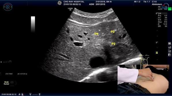
What the Liver Looks Like on Abdominal Ultrasound
The liver is the largest solid organ in the abdomen and one of the primary targets of abdominal ultrasound. A normal liver has a homogeneous medium-grey echotexture on ultrasound, smooth regular margins, and well-visualised hepatic veins and portal veins. Several important liver conditions produce characteristic ultrasound appearances that can be identified and reported by a radiologist. Hepatic steatosis (fatty liver) produces increased echogenicity — the liver appears brighter than normal, and brighter than the right kidney (which is used as a reference structure). Severe fatty infiltration produces a “bright liver” appearance with poor visualisation of the portal vein walls and diaphragm due to sound attenuation. Ultrasound is sensitive for detecting moderate-to-severe hepatic steatosis (sensitivity approximately 80–90% for steatosis above 20–30%) but less reliable for mild steatosis or quantification, which is where FibroScan (controlled attenuation parameter, CAP) and MRI-PDFF add precision. Liver cirrhosis produces a coarsened echotexture — the liver parenchyma loses its smooth homogeneous appearance and becomes irregular and nodular. Other cirrhosis findings include irregular liver margins, right lobe atrophy with caudate lobe hypertrophy, splenomegaly (an enlarged spleen reflecting portal hypertension), ascites (free fluid in the peritoneal cavity), and portal vein dilation. While ultrasound can suggest cirrhosis through these findings, it cannot histologically grade fibrosis — liver biopsy or elastography (FibroScan) provides the objective fibrosis quantification covered in the dedicated article on liver function tests. Liver masses — haemangiomas (common benign vascular tumours producing a well-defined hyperechoic nodule), liver cysts (smooth thin-walled anechoic lesions with posterior acoustic enhancement), hepatocellular carcinoma (HCC — variable appearance, often hypo- or hyperechoic nodules in a cirrhotic liver), and metastases (multiple lesions of variable echogenicity) — are detected by ultrasound, though characterisation of indeterminate liver lesions often requires CT or MRI with contrast for definitive diagnosis.

Gallbladder and Bile Ducts on Abdominal Ultrasound
The gallbladder and bile ducts are ideally suited to ultrasound assessment — the gallbladder is a fluid-filled sac that provides a natural acoustic window, and ultrasound is the most accurate imaging modality for detecting gallstones (sensitivity 95–99%, specificity 99%). Gallstones (cholelithiasis) appear as bright (hyperechoic) foci within the gallbladder lumen that cast a posterior acoustic shadow (a dark region behind the stone caused by sound attenuation through the calcified stone). Gallstones are typically mobile — they move to the dependent portion of the gallbladder with patient repositioning — distinguishing them from gallbladder polyps (which are fixed). Small stones or sludge (suspended particulate bile material) may not cast a shadow and can be missed in a poorly prepared (non-fasted) patient. Acute cholecystitis (gallbladder inflammation, most commonly caused by a stone obstructing the cystic duct) produces characteristic ultrasound findings: gallbladder wall thickening above 3 mm, pericholecystic fluid (fluid around the gallbladder), and the sonographic Murphy’s sign — maximum tenderness directly over the gallbladder when the transducer is pressed on it. A positive sonographic Murphy’s sign in the context of gallbladder wall thickening is highly predictive of acute cholecystitis. Common bile duct dilation (diameter above 7 mm in patients without a prior cholecystectomy) is an important finding suggesting biliary obstruction — from common bile duct stones (choledocholithiasis), stricture, or pancreatic head mass. The common bile duct is measured on ultrasound to screen for obstruction, though the stones themselves may not be visualised (gas in the duodenum adjacent to the common bile duct is a common ultrasound limitation). When bile duct dilation is identified, further imaging with MRCP (magnetic resonance cholangiopancreatography) is typically the next step to characterise the level and cause of obstruction. The symptom of pale stool from biliary obstruction — covered in the article on pale stool and liver or bile problems — is one of the clinical triggers prompting urgent abdominal ultrasound.
Kidneys, Spleen, Pancreas, and Aorta
Beyond the liver and gallbladder, abdominal ultrasound provides important information about the kidneys, spleen, pancreas, and aorta. Kidney assessment includes measurement of kidney size (normal 9–12 cm in length), evaluation for hydronephrosis (dilation of the renal collecting system from urinary tract obstruction), detection of kidney stones (appearing as bright foci with acoustic shadow), assessment of kidney cysts (common incidental findings, most of which are benign simple cysts), and evaluation of kidney parenchymal echogenicity (increased echogenicity suggests chronic kidney disease). Hydronephrosis — dilation of the renal pelvis and calyces from obstructed urine flow — is graded from mild to severe and may require urgent urology referral if bilateral or causing acute kidney injury. Spleen assessment measures splenic size (splenomegaly — defined as a spleen above 12 cm in length — is an important finding suggesting portal hypertension from liver cirrhosis, haematological malignancy, or chronic infection) and evaluates for splenic lesions. Pancreas assessment is one of the most technically challenging aspects of abdominal ultrasound because the pancreas lies retroperitoneally behind the stomach and overlying bowel gas frequently obscures it. When visible, ultrasound can assess pancreatic size, detect pancreatic duct dilation (above 3 mm — a sign of chronic pancreatitis or pancreatic head obstruction), and identify pancreatic masses. However, CT or MRI is more reliable for pancreatic pathology when ultrasound is technically limited. Abdominal aorta measurement — the screening measurement for abdominal aortic aneurysm (AAA) — is included in most abdominal ultrasounds; an aortic diameter above 3 cm is considered dilated and above 5.5 cm (or above 5.0 cm in women) warrants surgical assessment. AAA screening by ultrasound in men over 65 is one of the most evidence-based screening programmes in vascular medicine. The relationship between abdominal findings on ultrasound and the biochemical markers of liver disease — including ALT, AST, and GGT — is covered in the article on ALT and AST blood tests and the GGT test.
Limitations of Abdominal Ultrasound
Understanding what abdominal ultrasound cannot reliably detect is as clinically important as understanding what it can show — patient expectations and clinical decision-making both depend on this knowledge. The primary limitations of abdominal ultrasound include: operator and patient dependence — ultrasound image quality depends significantly on the sonographer’s skill and experience, the ultrasound machine quality, and patient factors including body habitus (abdominal fat attenuates sound waves, reducing image quality in obese patients) and bowel gas content. Bowel visualisation — gas-containing bowel reflects ultrasound, making the small intestine and colon largely inaccessible to ultrasound in most patients. Bowel pathology including colorectal cancer, intestinal Crohn’s disease, and small bowel obstruction are therefore poorly assessed by standard abdominal ultrasound. Retroperitoneal structures — the pancreas, retroperitoneal lymph nodes, and retroperitoneal masses are often obscured by overlying bowel gas, making CT or MRI the preferred modality when retroperitoneal pathology is suspected. Characterisation of liver lesions — while ultrasound detects focal liver lesions, it often cannot reliably characterise them as benign or malignant without contrast enhancement. A lesion found on ultrasound that cannot be definitively characterised is an indication for CT or MRI with contrast to provide more definitive tissue characterisation. Depth limitation — deep structures in obese patients may be beyond the effective penetration depth of the transducer, producing non-diagnostic images. In these situations, contrast-enhanced ultrasound (CEUS), CT, or MRI provides superior anatomical coverage. Understanding when abdominal ultrasound findings need to be followed up with more advanced imaging — CT scan covered in a separate article — is an important part of the clinical decision-making that follows an ultrasound report. The role of dark urine, jaundice, and abdominal swelling as clinical triggers for urgent imaging is explored in the articles on dark urine and liver health and swollen belly and liver disease.
Frequently Asked Questions About Abdominal Ultrasound
How do I prepare for an abdominal ultrasound?
Most abdominal ultrasound requests require fasting for four to six hours before the scan. The primary reason for fasting is to ensure the gallbladder is distended with bile — when the gallbladder is well-filled, gallstones are more visible and wall assessment is more reliable. Eating causes the gallbladder to contract, expelling bile and making it small and contracted, which reduces sensitivity for gallstone detection. Fasting also reduces bowel gas, slightly improving visualisation of retroperitoneal structures. Water is generally permitted during the fasting period as it does not stimulate gallbladder contraction. Some ultrasound departments request patients to arrive with a full bladder (drinking 500–750 ml of water one hour before the scan) if pelvic structures (bladder, uterus, ovaries in a female pelvis ultrasound) are also being assessed. Before the scan, patients change into a hospital gown, lie on an examination table, and a gel (acoustic coupling gel, which looks and feels similar to hair gel) is applied to the abdominal skin. The transducer is pressed against the skin and moved across the abdomen to obtain images. Patients may be asked to take deep breaths to lower the diaphragm and bring the upper abdominal organs into better view. The scan typically takes twenty to forty minutes depending on the number of organs assessed and the technical difficulty.
My ultrasound showed a “bright liver” — what does this mean?
A “bright liver” or “increased hepatic echogenicity” on ultrasound is the most common radiological report finding associated with hepatic steatosis — fatty liver disease. The finding describes the liver appearing more echogenic (brighter) than normal on the ultrasound image, reflecting increased fat content in hepatocytes causing more sound wave reflection. This is a non-specific finding — while hepatic steatosis is by far the most common cause, increased liver echogenicity can also result from fibrosis, cirrhosis, glycogen storage disease, and other parenchymal conditions. A “bright liver” finding should prompt further clinical evaluation: blood tests including liver function tests (ALT, AST, GGT, bilirubin, albumin), metabolic risk factor assessment (fasting glucose, HbA1c, lipids, BMI), and consideration of FibroScan to assess coexistent fibrosis, particularly when ALT is elevated or metabolic risk factors are present. Hepatic steatosis identified on ultrasound is a modifiable condition — lifestyle intervention targeting weight loss, regular physical activity, and dietary changes (reducing ultra-processed foods, refined carbohydrates, and alcohol) can significantly reduce hepatic fat content and improve ultrasound findings on follow-up. The biochemical test context for fatty liver — including the liver enzyme patterns covered in alkaline phosphatase testing — provides the laboratory framework alongside ultrasound findings.
Sources: ACR Appropriateness Criteria — Abdominal Imaging · NIDDK — Abdominal Ultrasound · RadiologyInfo.org — Abdominal Ultrasound
Ascites Detection and Portal Hypertension Assessment
Ascites — the accumulation of free fluid in the peritoneal cavity — is one of the most important findings detectable by abdominal ultrasound, and ultrasound is the most sensitive technique for detecting even small volumes of abdominal free fluid that are not clinically apparent on physical examination. As little as 100 ml of peritoneal fluid can be detected by ultrasound, compared with approximately 1500 ml required for clinical detection by abdominal percussion and shifting dullness. Ascites appears on ultrasound as anechoic (dark) fluid in dependent abdominal spaces — the hepatorenal space (Morison’s pouch), the splenorenal space, the paracolic gutters, and the pelvis. Simple transudative ascites (from liver cirrhosis, heart failure, or hypoalbuminaemia) is anechoic and featureless. Exudative ascites (from malignancy, spontaneous bacterial peritonitis, tuberculosis) may contain echogenic debris, septations, or loculations. Ultrasound-guided ascitic tap (paracentesis) — draining the ascitic fluid for diagnostic analysis or therapeutic relief — uses real-time ultrasound to identify the safest needle entry site, avoiding bowel and vascular injury. Portal hypertension — the increased pressure in the portal venous system from cirrhosis or portal vein thrombosis — is assessed indirectly on ultrasound through several findings: portal vein dilation (greater than 13 mm), splenomegaly (spleen above 12 cm), ascites, and the presence of portosystemic collateral vessels (including splenorenal shunts and recanalized paraumbilical veins). Doppler ultrasound — which assesses blood flow velocity and direction using the Doppler effect of sound waves in moving blood — can directly measure portal vein flow velocity and detect portal vein thrombosis (absent or reversed flow). In patients with known liver cirrhosis, regular ultrasound surveillance every six months is recommended to screen for hepatocellular carcinoma (HCC) — the most important complication of cirrhosis — with AFP (alpha-fetoprotein) blood test performed simultaneously. This surveillance programme is endorsed by EASL and AASLD guidelines. The visible consequences of liver disease including jaundice (covered in the article on yellow skin or eyes) and ascites provide the clinical context within which urgent abdominal ultrasound is ordered.
Doppler Ultrasound and Vascular Assessment
Doppler ultrasound extends standard B-mode (greyscale) ultrasound by adding the ability to assess blood flow using the Doppler frequency shift — the change in frequency of sound waves reflected by moving red blood cells. Colour Doppler ultrasound overlays flow information onto the B-mode image: blood flowing towards the transducer appears red; blood flowing away appears blue (by convention). Spectral Doppler measures flow velocity over time, producing a waveform that characterises the flow pattern in vessels. In the abdominal context, Doppler ultrasound is used for: portal vein assessment (measuring velocity, detecting thrombosis, identifying flow reversal in portal hypertension); hepatic artery assessment (important post liver transplantation to confirm arterial patency); hepatic vein waveform assessment (the Budd-Chiari syndrome — hepatic venous outflow obstruction — produces absent or reversed hepatic vein flow); renal artery Doppler (assessing for renal artery stenosis as a cause of hypertension or renal impairment); and abdominal aorta assessment (measuring aortic diameter for AAA surveillance and characterising aortic dissection). Duplex ultrasound combines B-mode and Doppler in a single examination, providing both anatomical and flow information simultaneously. The combination of greyscale abdominal ultrasound and Doppler assessment is a comprehensive non-invasive evaluation of the abdominal vascular and solid organ anatomy that guides the need for more advanced imaging modalities including CT and MRI. The article on easy bruising and liver function covers the coagulopathic consequences of portal hypertension that the vascular Doppler findings on ultrasound help explain.
When Abdominal Ultrasound Findings Need Further Investigation
Not every abdominal ultrasound finding is diagnostic in isolation — many findings require correlation with clinical symptoms, blood test results, and often further imaging to reach a definitive clinical conclusion. Understanding when an ultrasound report warrants urgent follow-up versus routine monitoring is important for patients interpreting their results. Findings that typically require urgent further assessment include: focal liver lesions in patients with cirrhosis or known malignancy (require contrast-enhanced CT or MRI within days to weeks); dilated bile duct above 10 mm (requires urgent MRCP or endoscopic assessment to exclude malignancy); hydronephrosis with urosepsis features (requires urgent urology assessment); aortic diameter approaching surgical threshold; and new large-volume ascites without known cause (requires urgent paracentesis and investigation for malignancy or infection). Findings that warrant routine follow-up include: indeterminate liver cysts above 1 cm (characterisation by CT or MRI); simple kidney cysts requiring periodic size monitoring; mildly dilated bile duct in an older patient with prior cholecystectomy (requires correlation with liver blood tests and symptoms). A “negative” or “normal” abdominal ultrasound does not exclude all pathology — bowel disease, early pancreatic cancer, and retroperitoneal lymphadenopathy may not be visualised. Clinical symptoms that persist despite a normal ultrasound are an indication for CT or endoscopic investigation. The full diagnostic context of imaging findings alongside blood tests — including the bilirubin, albumin, and alkaline phosphatase covered in the articles on bilirubin testing and albumin and liver function — provides the integrated clinical picture that guides management decisions.
Abdominal ultrasound is a fundamental first-line diagnostic tool that provides a remarkable amount of clinical information from a rapid, safe, and widely available investigation. It is the starting point for imaging the liver, gallbladder, kidneys, spleen, and major vessels in the vast majority of clinical presentations — from right upper quadrant pain and jaundice to unexplained weight loss and abnormal blood tests. Its limitations — operator dependence, bowel gas interference, limited retroperitoneal access — are well understood and define the indications for escalation to CT or MRI rather than representing a reason to bypass ultrasound as first-line investigation. For patients receiving abdominal ultrasound reports, the key is to understand that each finding — from a bright liver to a dilated bile duct to a simple kidney cyst — has a defined clinical significance and a defined management pathway that your clinician will use to guide next steps. The connection between ultrasound findings and the blood tests that provide the biochemical counterpart to the imaging picture — liver function tests, bilirubin, albumin, GGT — is explored throughout the articles on liver function tests explained and the hepatitis blood tests series, providing the integrated clinical context that turns individual test results into actionable diagnostic conclusions.
The evolution of point-of-care ultrasound (POCUS) — bedside ultrasound performed by the treating clinician rather than a dedicated radiologist — has expanded the role of ultrasound in emergency and acute care settings. POCUS abdominal assessments in emergency medicine focus on targeted questions: Is there free fluid in the abdomen? Is the aorta dilated? Is the gallbladder inflamed? Is there hydronephrosis? These focused assessments can be performed within minutes at the bedside, guiding immediate management decisions without the delay of formal radiology. Understanding what abdominal ultrasound can show — in its full formal and point-of-care applications — equips patients to understand why this investigation is ordered both as routine outpatient assessment and as urgent bedside evaluation in acute presentations.
My GP ordered an abdominal ultrasound after I had elevated liver enzymes on a routine blood test — I had no symptoms at all. The scan showed a bright liver consistent with fatty liver and a 6mm gallstone. I had no idea either of those things was there. The radiologist’s report was quite technical but this article helped me understand exactly what ‘increased echogenicity’ and ‘posterior acoustic shadowing’ actually meant.
Karen, a ‘bright liver’ finding alongside elevated ALT is a common and important combination — the ultrasound confirms fatty infiltration while the blood tests tell you it is causing some degree of hepatocellular stress. A 6mm gallstone without symptoms is generally managed conservatively (watchful waiting), as the risk of complications from small asymptomatic stones is low. The key next step is understanding your metabolic risk factors for fatty liver disease — weight, blood glucose, lipids, alcohol — since addressing these can lead to meaningful improvement in both the liver enzyme picture and the ultrasound appearance over time.
I just want to highlight the AAA screening point — my father had a 5.2cm aortic aneurysm found on a routine abdominal ultrasound before it ruptured. He had surgery and is fine. The NHS screening programme for men at 65 is genuinely life-saving and I’d encourage every man in that age group to attend when invited. An ultrasound scan of the aorta takes about five minutes.