
Magnetic resonance imaging (MRI) of the liver uses a powerful magnetic field and radiofrequency pulses — rather than ionising radiation — to generate highly detailed cross-sectional images of the liver parenchyma, focal lesions, biliary system, and hepatic vasculature. In liver medicine, MRI has become the definitive characterisation tool for focal liver lesions that are indeterminate on ultrasound or CT, the most accurate modality for biliary tract evaluation (via MRCP — magnetic resonance cholangiopancreatography), and — with liver-specific contrast agents — a functional imaging tool that assesses hepatocellular uptake capacity alongside anatomical lesion characterisation. Understanding when an MRI is ordered for liver evaluation, what each component of the MRI examination assesses, what the results mean for different liver conditions, and what to expect during the procedure allows patients to engage with this important investigation with confidence and clarity.
MRI works by placing the patient in a strong magnetic field (typically 1.5 or 3 Tesla) that aligns hydrogen nuclei (protons) in the body. Radiofrequency pulses perturb these protons, and as they return to their equilibrium state they emit signals that are detected and processed by computer into images. Unlike CT, MRI does not use ionising radiation — this is its principal safety advantage, making it the preferred modality for repeated imaging, for younger patients, and for pregnant women (after the first trimester, when indicated). Different tissue types (liver parenchyma, fat, blood, tumour) return different MRI signal intensities on different pulse sequences, providing exceptional soft tissue contrast that exceeds what CT can offer. The specific MRI sequences used for liver evaluation — T1-weighted (which shows fat content and detects haemorrhage), T2-weighted (which shows fluid and is sensitive for cysts and certain tumours), diffusion-weighted imaging (DWI — which reflects tissue cellularity and is highly sensitive for detecting liver lesions including small metastases and HCC), and dynamic contrast-enhanced sequences (using intravenous gadolinium-based contrast) — together provide a comprehensive, multi-dimensional assessment of liver structure and pathology.
Liver MRI for Focal Lesion Characterisation
The primary clinical indication for liver MRI is characterisation of focal liver lesions that are indeterminate on ultrasound or CT. Liver MRI provides superior soft tissue contrast and access to multiple complementary imaging sequences that together allow definitive characterisation of most liver lesions without biopsy. Hepatocellular carcinoma (HCC) is diagnosed on MRI by the LI-RADS (Liver Imaging Reporting and Data System) classification, which categorises liver observations from LR-1 (definitely benign) to LR-5 (definitely HCC) based on imaging features including: arterial phase hyperenhancement (APHE — the lesion enhances more than surrounding liver in the arterial phase, reflecting HCC’s arterial blood supply); washout appearance (the lesion becomes darker than surrounding liver in the portal venous or delayed phase, reflecting washout of contrast); enhancing capsule (a visible rim of enhancement around the lesion, reflecting the fibrous capsule of HCC); and size above 10 mm. An LR-5 observation on MRI with liver-specific contrast (gadoxetate) in a cirrhotic patient is diagnostic for HCC, enabling treatment planning without tissue biopsy. Liver metastases are detected with high sensitivity on DWI sequences (appearing as bright foci) and characterised by their enhancement pattern — colorectal metastases are hypovascular; neuroendocrine metastases are hypervascular. MRI is more sensitive than CT for detecting small hepatic metastases (less than 1 cm), making it the preferred modality for pre-operative metastasis staging before liver surgery. Benign liver lesions — haemangiomas (characteristic T2 bright signal and progressive peripheral enhancement), simple cysts (uniform T2 bright, no enhancement), focal nodular hyperplasia (FNH — T1 isointense with central scar, early enhancement, retention of gadoxetate in the hepatobiliary phase), and hepatic adenoma (fat signal on chemical shift imaging, variable enhancement) — are characterised on MRI by their specific combination of T1, T2, diffusion, and enhancement features. For indeterminate liver lesions found on ultrasound or CT, MRI resolves the diagnostic uncertainty in the majority of cases, directing appropriate management — surveillance, biopsy, or treatment. The blood test context for liver lesion assessment — including AFP as an HCC marker alongside the liver function tests covered in the article on liver function tests explained — provides the biochemical picture alongside MRI findings.
Liver-Specific MRI Contrast — Gadoxetate (Primovist/Eovist)
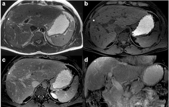
Gadoxetate (sold as Primovist in Europe/Asia and Eovist in North America) is a liver-specific MRI contrast agent that provides both standard dynamic vascular phases (arterial, portal venous) and a unique hepatobiliary phase (HBP) acquired twenty minutes after injection, when functioning hepatocytes take up the contrast agent. This hepatobiliary phase provides functional information about hepatocyte uptake capacity that is unavailable with standard extracellular gadolinium-based contrast: Normal hepatocytes take up gadoxetate, so normal liver parenchyma appears bright on the HBP. HCC — which consists of abnormal tumour cells with reduced hepatocyte organic anion transporter (OATP) expression — does not take up gadoxetate and appears dark (non-enhancing) on the HBP, creating the “washout” appearance that is one of the LI-RADS diagnostic criteria. FNH — which contains functioning hepatocytes — takes up gadoxetate and appears iso- or hyperintense on the HBP, an important distinguishing feature from hepatic adenoma (which does not enhance on HBP). Cholangiocarcinoma — a biliary tumour — shows peripheral enhancement and delayed central fill-in; it does not take up gadoxetate on the HBP. The hepatobiliary phase also provides visualisation of the biliary anatomy, allowing assessment of bile duct patency and biliary communications. Gadoxetate MRI is the most comprehensive and diagnostically accurate liver imaging examination available, combining dynamic vascular characterisation with functional hepatocyte assessment and biliary information in a single investigation. Its use is particularly important in: pre-operative planning before liver resection or transplantation (the HBP provides functional mapping of which liver segments retain function, guiding the extent of safe resection); indeterminate liver lesions in cirrhosis (LI-RADS 3 observations that require follow-up characterisation); and post-treatment response assessment after locoregional therapy (ablation, TACE). The connection between hepatic functional capacity — reflected by gadoxetate HBP uptake — and the synthetic liver blood tests covered in the article on albumin and liver function provides the integrated picture of hepatic reserve assessment.
MRCP — MRI of the Biliary System
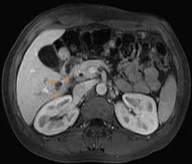
Magnetic resonance cholangiopancreatography (MRCP) is a non-invasive MRI technique that produces heavily T2-weighted images in which fluid-containing structures — bile ducts and the pancreatic duct — appear bright, creating a three-dimensional roadmap of the biliary tree and pancreatic ductal anatomy. MRCP has replaced diagnostic ERCP (endoscopic retrograde cholangiopancreatography) for the majority of biliary diagnostic indications because it provides equivalent anatomical information without the risks of ERCP (acute pancreatitis in approximately three to five percent, bile duct perforation, cholangitis). ERCP is now reserved for therapeutic indications — stone extraction, biliary stenting, tissue sampling — where endoscopic access is required for treatment in addition to diagnosis. Clinical indications for MRCP include: suspected common bile duct stones (choledocholithiasis) causing biliary obstruction (jaundice, right upper quadrant pain, elevated bilirubin and ALP); evaluation of biliary strictures (distinguishing benign from malignant strictures, characterising the level of obstruction); primary sclerosing cholangitis (PSC — MRCP characterises the multifocal biliary stricturing pattern of this autoimmune bile duct disease, monitors for dominant stricture development and cholangiocarcinoma); pancreatic ductal abnormalities including IPMN (intraductal papillary mucinous neoplasm — an increasingly detected cystic pancreatic lesion requiring surveillance for malignant transformation); and pre-operative biliary anatomy mapping before liver or pancreatic surgery. The relationship between biliary obstruction and the specific blood test pattern it produces — elevated bilirubin and ALP covered in the articles on bilirubin testing and alkaline phosphatase — provides the clinical trigger for ordering MRCP in a jaundiced patient.