
Nonalcoholic fatty liver disease — widely known by its abbreviation NAFLD — was for decades the standard clinical term for fatty liver disease caused by metabolic dysfunction rather than alcohol consumption. The term distinguished this increasingly common condition from alcohol-related liver disease and gave clinicians a consistent label for what is now understood to be the most common liver disease in the world. In 2023, an international expert consensus led by leading liver disease societies formally retired the NAFLD label and replaced it with metabolic dysfunction-associated steatotic liver disease (MASLD) — a change that more accurately reflects the metabolic origins of the condition and removes a name that defined a disease primarily by the absence of something (alcohol) rather than by its actual cause. Despite this renaming, NAFLD remains widely used in older medical literature, patient information materials, and GP consultation notes, meaning that patients encountering the term today — in blood test reports, referral letters, or historical records — need to understand both labels and their relationship to current terminology.
This article explains what NAFLD was, how it relates to the current MASLD diagnosis, what the condition involves at a biological and clinical level, how it is discovered and diagnosed, how it progresses, what determines prognosis, and what the current approach to management entails. Whether you have received a letter using the NAFLD label or have recently been told you have MASLD — or are simply trying to understand the condition in the context of a fatty liver finding on ultrasound — this guide provides the foundational understanding needed to engage with the investigation and management process.
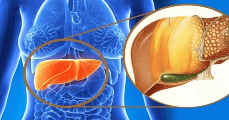
NAFLD and the 2023 Terminology Change
The NAFLD label was introduced in 1980 to describe a pattern of liver disease that resembled alcoholic liver disease histologically — the same fat accumulation, the same hepatocyte injury, the same fibrosis — but occurring in patients with little or no alcohol consumption. The name was useful in distinguishing metabolic liver disease from alcohol-related disease at a time when the distinction was not well understood, but over time its limitations became apparent. First, it defined the condition by what it was not (non-alcoholic) rather than by its actual mechanism, which created confusion for patients and clinicians. Second, it carried implicit stigma by associating the condition with alcohol use through negation. Third, the original NAFLD definition required the explicit exclusion of alcohol as a contributing factor, which created a false binary: many patients have both metabolic dysfunction and some level of alcohol use, neither of which adequately explains the totality of their liver disease under the old framework. The 2023 consensus — published by the EASL, AASLD, and their partner societies — replaced NAFLD with MASLD and NASH (nonalcoholic steatohepatitis) with MASH (metabolic dysfunction-associated steatohepatitis). The new nomenclature defines the condition by its metabolic cause rather than by the absence of alcohol and introduces a separate category, MetALD, for patients with metabolic dysfunction who also drink at levels that could be contributing to their liver disease. For practical purposes, NAFLD = MASLD and NASH = MASH in the vast majority of cases. Patients who received a NAFLD diagnosis before 2023 have the same condition as patients now receiving an MASLD diagnosis.
What NAFLD/MASLD Actually Is
At its core, MASLD is a disease of impaired hepatic fat metabolism caused by metabolic dysfunction — specifically insulin resistance. In a metabolically healthy liver, lipid metabolism is tightly regulated: fatty acids arriving at the liver (from dietary fat and from adipose tissue lipolysis) are balanced against fatty acid oxidation (burning fat for energy), export of lipids as VLDL particles, and de novo lipogenesis (synthesis of new fat from carbohydrates). In insulin resistance — the metabolic state that underlies MASLD — this balance is disrupted: adipose tissue releases more free fatty acids into the portal circulation, the liver receives a greater lipid load, insulin resistance impairs normal fatty acid oxidation and lipid export, and de novo lipogenesis is paradoxically increased despite high insulin levels. The result is net fat accumulation within hepatocytes — the liver cells that constitute the functioning liver parenchyma. When more than five percent of hepatocytes contain visible fat droplets (steatosis), the histological and clinical definition of fatty liver is met. This accumulation of fat is not inherently toxic to hepatocytes at low levels — simple steatosis is largely benign — but the metabolic environment that produces it (insulin resistance, oxidative stress, adipokine imbalance, gut microbiome disruption) also creates the conditions for ongoing hepatocyte injury, and in a subset of patients, this progresses to the more severe form of the disease: steatohepatitis (MASH in current terminology, NASH under the old NAFLD framework). The condition is therefore best understood not as a liver disease that happens to occur in people with metabolic syndrome, but as a hepatic manifestation of metabolic syndrome itself — the liver’s response to the same pathological metabolic state that drives type 2 diabetes, cardiovascular disease, and hypertension.
NAFLD Versus NASH: Understanding the Spectrum
The original NAFLD label covered a spectrum of disease severity that was divided into two major categories: simple NAFLD (now: simple steatosis within MASLD) and NASH (now: MASH). Understanding this distinction remains important because it determines prognosis and management intensity.
Simple steatosis (MASLD without steatohepatitis): Fat accumulation in more than five percent of hepatocytes, without significant liver cell damage or inflammation. Liver blood tests may be mildly elevated or normal. Histologically, the liver shows fat but no hepatocyte ballooning (a specific form of cellular injury), minimal inflammation, and no or minimal fibrosis. Simple steatosis is the most common form of MASLD — present in approximately seventy-five percent of patients with MASLD — and carries a relatively benign prognosis: the risk of progression to advanced fibrosis over a ten-year period is approximately ten to twenty percent. Most patients with simple steatosis will not develop cirrhosis, and the fat accumulation is reversible with lifestyle change. Steatohepatitis (MASH, formerly NASH): The more severe, progressive form of MASLD, characterised by steatosis combined with hepatocyte ballooning, lobular inflammation, and often early fibrosis. Present in approximately twenty-five percent of MASLD patients. MASH represents active liver disease — the liver is being damaged on an ongoing basis — and carries a significantly higher risk of fibrosis progression, cirrhosis, liver failure, and hepatocellular carcinoma than simple steatosis. Distinguishing MASH from simple steatosis cannot be done reliably on blood tests or non-invasive imaging alone — liver biopsy has historically been the only way to make the histological diagnosis — though various serum fibrosis indices and imaging technologies are increasingly used to identify high-risk patients without biopsy. The activity and fibrosis scores used in liver biopsy assessment (NAS score: Non-alcoholic Fatty Liver Disease Activity Score, which retains its old acronym even under new terminology) grade the severity of steatohepatitis and fibrosis, informing both prognosis and clinical trial eligibility.

How NAFLD/MASLD Is Discovered
Like most liver diseases, NAFLD/MASLD is usually asymptomatic — it causes no symptoms that would prompt a patient to seek medical attention until the disease is at an advanced stage (significant fibrosis or cirrhosis). The vast majority of diagnoses are made incidentally, through one of three routes:
Abnormal liver blood tests on routine testing: ALT and AST elevation is the most common pathway — a mildly elevated ALT (often one to three times the upper limit of normal) discovered during routine health checks, pre-operative assessment, medication monitoring, or investigation of unrelated symptoms. GGT may also be elevated, particularly in patients who drink any alcohol. The full workup of abnormal liver function tests proceeds through exclusion of other liver diseases before MASLD is confirmed as the diagnosis. Incidental finding on ultrasound: An abdominal ultrasound performed for gallstones, right upper quadrant pain, or other indications may show a bright (echogenic) liver — the sonographic appearance of hepatic steatosis. An echogenic liver does not confirm MASLD or quantify fibrosis; it is a prompt for further assessment. Metabolic disease monitoring: Patients under active management for obesity, type 2 diabetes, or metabolic syndrome may have liver tests checked as part of routine monitoring, identifying MASLD as a hepatic complication of their underlying metabolic disease before it causes any liver-specific symptoms. In each of these discovery routes, the initial finding prompts the same investigation pathway: establishing the aetiology (confirming MASLD by excluding other liver diseases) and staging the severity (quantifying fibrosis to determine management intensity and risk).
The Diagnosis and Investigation of NAFLD/MASLD
The diagnosis of MASLD is clinical — it requires the presence of hepatic steatosis (on imaging or biopsy) plus at least one cardiometabolic risk factor (overweight/obesity, impaired glucose tolerance or type 2 diabetes, hypertension, raised triglycerides, low HDL cholesterol) and the exclusion of significant alcohol consumption and other liver diseases. The investigation pathway that follows a fatty liver finding encompasses several elements:
Excluding other liver diseases: Blood tests assess hepatitis B and C serology, autoimmune liver markers (ANA, SMA, AMA), thyroid function, ferritin and transferrin saturation, alpha-1 antitrypsin, and in younger patients, Wilson’s disease markers. Medication review excludes drug-induced liver disease. A systematic alcohol history is taken. This exclusion panel is essential because MASLD is a diagnosis of inclusion (steatosis plus metabolic risk factor) as well as exclusion (ruling out other causes of elevated enzymes and hepatic steatosis). Fibrosis staging with the FIB-4 index: The FIB-4 score (calculated from age, ALT, AST, and platelet count) is used as a first-line triage tool in primary care. A FIB-4 below 1.30 has a high negative predictive value for advanced fibrosis, effectively identifying patients in the low-risk category who can be monitored in primary care. A FIB-4 above 2.67 indicates high probability of advanced fibrosis and warrants specialist hepatology referral and further assessment. Patients in the intermediate range (1.30–2.67) benefit from FibroScan for further characterisation. FibroScan: Vibration-controlled transient elastography measures liver stiffness (in kPa) as a proxy for fibrosis stage, and the CAP score quantifies hepatic steatosis grade (S0–S3) using the attenuation of the ultrasound signal. The FibroScan guide on this site explains the procedure, its interpretation, and its limitations in detail. FibroScan is now the primary non-invasive fibrosis staging tool in the NAFLD/MASLD pathway, recommended in all major clinical guidelines as the first-line modality following FIB-4 risk stratification. Liver biopsy: Reserved for cases where non-invasive tests are discordant, where clinical decision-making requires histological confirmation (for example, to diagnose MASH and quantify activity before pharmacological treatment), or where another liver disease cannot be excluded non-invasively. The procedure, its indications, risks, and what it shows are covered in the dedicated liver biopsy article in this series.
Prognosis: Who Progresses and Who Does Not
One of the most important and often misunderstood aspects of NAFLD/MASLD is its heterogeneous natural history — most patients with MASLD do not progress to cirrhosis, but a meaningful minority do, and identifying which patients are in that minority is the central challenge of MASLD management. The strongest predictor of liver-related outcomes is fibrosis stage — specifically, whether a patient has advanced fibrosis (F3–F4) or not. Patients with F0–F1 fibrosis (no fibrosis or mild fibrosis) have a liver-related mortality risk that is not significantly elevated above the general population. Patients with F3–F4 fibrosis have substantially elevated risks of liver-related complications, hepatocellular carcinoma, and liver-related death, even after adjusting for other causes of death. In the largest published meta-analyses, liver-related mortality increases in an approximately stepwise fashion with fibrosis stage: patients with F4 (cirrhosis) have roughly a ten-fold higher liver-related mortality risk than those with F0. The presence of MASH (steatohepatitis) at the histological level predicts faster fibrosis progression than simple steatosis, independently of current fibrosis stage — a patient with F1 fibrosis and MASH is at greater risk of progressing to F3 than a patient with F1 fibrosis and simple steatosis. Metabolic risk factor control is a major modifiable determinant of progression trajectory: patients who achieve sustained weight loss, improve glycaemic control, and reduce dyslipidaemia show documented fibrosis regression on serial FibroScan and biopsy assessments. The EWAS and LEAN trials, and observational data from bariatric surgery cohorts, provide compelling evidence that metabolic improvement translates into histological improvement. The implication is that the prognosis of NAFLD/MASLD is not fixed at diagnosis — it is determined by a combination of baseline fibrosis stage and subsequent metabolic risk factor management, which means that a fatty liver diagnosis, taken seriously and managed actively, carries a substantially better long-term prognosis than one that is ignored.
Management of NAFLD/MASLD
The treatment of MASLD rests on three pillars: lifestyle modification, metabolic risk factor management, and — for patients with MASH and significant fibrosis — increasingly, pharmacological therapy targeted at the liver itself. Lifestyle modification: Weight loss is the most effective intervention for MASLD at every stage of disease. A five percent weight loss is associated with significant reduction in hepatic steatosis; a seven to ten percent loss is associated with histological improvement in MASH activity (inflammation and ballooning); a fifteen to twenty percent weight loss is associated with fibrosis regression. Sustained aerobic exercise independently reduces hepatic steatosis and improves insulin sensitivity, with benefits beyond what weight loss alone explains — current guidelines recommend one hundred and fifty minutes of moderate aerobic activity per week as a minimum target. Dietary pattern is also important: a Mediterranean-style diet (high in vegetables, whole grains, olive oil, fish, and legumes; low in refined carbohydrates and saturated fat) is associated with hepatic steatosis reduction independent of caloric restriction. Alcohol should be minimised even in MASLD (not ARLD) patients, as any alcohol intake appears to have an additive effect on liver injury in the context of existing steatosis. Metabolic risk factor management: Treating the underlying drivers of MASLD — glycaemic control in type 2 diabetes (with GLP-1 receptor agonists, SGLT2 inhibitors, or metformin), lipid management (statins are safe and beneficial in MASLD despite historical concerns), and blood pressure control — all contribute to reducing hepatic inflammation and fibrosis progression. Pharmacological therapy: Resmetirom, a thyroid hormone receptor-beta agonist, received FDA approval in March 2024 for the treatment of MASH with moderate to advanced fibrosis (F2–F3) — the first pharmacological agent specifically approved for MASH and representing a landmark in NAFLD/MASLD management. GLP-1 receptor agonists including semaglutide and liraglutide have demonstrated significant liver steatosis reduction in clinical trials and are in late-phase trials for MASH-specific approvals. Obeticholic acid and lanifibranor have shown fibrosis improvement in trials. The field of MASLD pharmacology has expanded significantly since 2020 and continues to evolve rapidly.
Frequently Asked Questions About NAFLD/MASLD
My doctor said I have NAFLD — do I now have MASLD?
Yes — NAFLD and MASLD refer to the same condition. The NAFLD label was retired in 2023 and replaced with MASLD following an international consensus. If you received a NAFLD diagnosis in previous years, you now have what is called MASLD in current clinical practice. The underlying condition, its causes, its spectrum, and its management approach are identical. The change is a terminological and conceptual update, not a change in the disease itself. The same applies to NASH — if you were diagnosed with NASH, the current equivalent is MASH. When you see references to NAFLD in older literature, referral letters, or online health information, you can treat them as equivalent to MASLD.
Is NAFLD/MASLD reversible?
Yes — particularly at the stage of simple steatosis, where the fat accumulation is entirely reversible with metabolic improvement. A ten percent reduction in body weight is consistently associated with significant reduction in liver fat as measured by FibroScan CAP score and MRI-PDFF. Even significant fibrosis (F2–F3) has been shown to regress with sustained lifestyle intervention, bariatric surgery, and pharmacological treatment. The evidence is clearest for bariatric surgery, where cohort studies with serial biopsies have documented cirrhosis regression in a subset of patients. The key point is that reversibility is not uniform across all stages: simple steatosis is highly reversible; MASH with early fibrosis is reversible with significant and sustained metabolic improvement; established cirrhosis is not fully reversible, though decompensated cirrhosis may convert to compensated cirrhosis with fibrosis regression. The implication is that the optimal time to intervene is before significant fibrosis develops — reinforcing the value of early fibrosis staging at diagnosis and active management of metabolic risk factors from that point forward.
Can I drink alcohol if I have NAFLD/MASLD?
Current guidelines advise that patients with MASLD should minimise alcohol consumption and aim to remain within national recommended limits at most, and ideally below them. Although MASLD is defined as a condition occurring without clinically significant alcohol use, any alcohol consumption adds a superimposed hepatic injury on top of the existing metabolic liver disease. Studies have shown that even moderate alcohol consumption accelerates fibrosis progression in patients with MASLD, and the hepatic steatosis and inflammation caused by MASLD and alcohol are additive rather than independent. Patients with MASLD who also drink above recommended limits have a condition now classified as MetALD under the updated 2023 nomenclature, which carries a higher risk than MASLD alone. The practical advice is: reducing or eliminating alcohol is one of the lower-effort, high-impact interventions available to patients with MASLD, and for patients who are not prepared to make significant dietary or activity changes, alcohol reduction is a valuable place to start.
Sources: EASL–EASD–EASO — MASLD Clinical Practice Guidelines · AASLD — Liver Disease Clinical Guidance · NIDDK — NAFLD and NASH
The Role of the Gut Microbiome in MASLD
Emerging evidence implicates gut microbiome dysbiosis as a contributing factor in MASLD development and progression — an area of active research that may yield additional therapeutic targets in the coming years. The gut and liver are connected by the portal circulation: bacteria-derived metabolites, lipopolysaccharides (LPS — components of gram-negative bacterial cell walls), short-chain fatty acids, and secondary bile acids produced in the gut travel directly to the liver via the portal vein. In patients with MASLD, gut microbiome composition is altered compared to metabolically healthy individuals — with reduced diversity and an increased ratio of Firmicutes to Bacteroidetes being among the most consistently reported findings, though the composition varies between studies. Increased intestinal permeability (“leaky gut”) in the setting of metabolic dysfunction allows LPS and bacterial products to enter the portal circulation in greater quantities, activating hepatic toll-like receptors (particularly TLR4) and triggering inflammatory signalling pathways that contribute to steatohepatitis. The specific gut microbiome profile associated with advanced fibrosis in MASLD has been characterised in several cohort studies, raising the possibility of microbiome-based fibrosis prediction. Interventions that modify the gut microbiome — including dietary modification, probiotics, prebiotics, and faecal microbiota transplantation — are under investigation in MASLD clinical trials, though none has yet achieved regulatory approval for this indication. The gut-liver axis represents a mechanistic dimension of MASLD that partially explains why dietary pattern (which shapes microbiome composition) influences liver disease activity independently of total caloric intake.
Really useful context on the name change from NAFLD to MASLD. My GP letter still says NAFLD and I wasn’t sure if I had a different condition to what’s mentioned online now. This cleared it up — it’s the same thing, just renamed. The section on reversibility was encouraging too.
Thank you Diane — the NAFLD to MASLD renaming has caused some understandable confusion, particularly because many GP systems and referral letters still use the old terminology. They refer to exactly the same condition, and any management advice or monitoring plan you received under the NAFLD label applies equally under MASLD. The key thing is to continue with the follow-up your GP recommended — FIB-4 calculation, FibroScan if indicated — so that your fibrosis stage is known and monitored. Glad the reversibility section was helpful: the evidence for that at the steatosis stage is really quite encouraging.
The explanation of how the gut microbiome connects to liver disease was something I hadn’t read about before in plain language. I have MASLD and IBS and it makes me wonder whether they’re related. I’ll raise the gut-liver axis topic with my gastroenterologist.