
A liver ultrasound is a targeted ultrasound examination focused specifically on the liver — its size, echotexture, margins, vasculature, and any focal or diffuse abnormalities. While the term “abdominal ultrasound” covers all organs in the abdomen, a “liver ultrasound” is directed by the clinical question specifically about the liver, often ordered when liver blood tests are abnormal or when a liver condition is already known and requires monitoring. Understanding when a liver ultrasound is clinically indicated, what it can show, how the findings are interpreted alongside liver blood tests, and what happens when ultrasound findings are uncertain provides patients with a comprehensive understanding of this commonly performed but sometimes incompletely understood investigation. The liver is one of the most sonographically accessible organs in the body — its large size, solid parenchyma, and superficial position in the right upper quadrant make it well-suited to ultrasound assessment, and the combination of liver ultrasound with liver function blood tests forms the cornerstone of non-invasive liver disease evaluation in both primary and secondary care.
Liver disease is often clinically silent in its early and intermediate stages — the liver has a large functional reserve and symptoms of liver dysfunction (fatigue, jaundice, ascites, bleeding) typically emerge only after substantial loss of functional liver parenchyma. This means that elevated liver enzymes on routine blood testing — the most common route to liver disease discovery — are often the first clue to a liver condition that would otherwise not be apparent clinically. When ALT, AST, or GGT are elevated on a blood test, one of the most important early investigations is a liver ultrasound: it can identify whether the parenchymal changes of fatty liver disease, fibrosis, or cirrhosis are present; whether there are focal lesions that require further characterisation; and whether the biliary system is dilated (suggesting obstruction) or whether portal hypertension is already established. The liver ultrasound findings, interpreted alongside the liver blood test pattern, guide the next steps of investigation including biopsy, FibroScan, or cross-sectional imaging with CT or MRI.
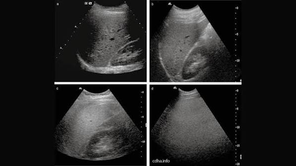
When a Liver Ultrasound Is Clinically Indicated
A liver ultrasound is indicated across a broad range of clinical situations, reflecting the central role of the liver in metabolism, detoxification, protein synthesis, and bile production. The most common indications include: Abnormal liver blood tests — elevated ALT, AST, GGT, alkaline phosphatase, or bilirubin on routine or targeted blood testing. Ultrasound is typically the first-line imaging investigation to evaluate the structural basis of biochemical liver abnormality. When ALT is elevated, ultrasound assesses for the hallmarks of fatty liver disease (the most common cause of elevated ALT in adults), viral hepatitis, autoimmune hepatitis, and less commonly focal hepatic pathology. The articles on ALT and AST blood tests and GGT testing provide the biochemical counterpart to liver ultrasound findings. Suspected fatty liver disease — patients with metabolic risk factors (obesity, type 2 diabetes, hypertriglyceridaemia, metabolic syndrome) or incidental finding of raised ALT undergo liver ultrasound to assess for hepatic steatosis. Known chronic liver disease monitoring — patients with confirmed MASLD (metabolic dysfunction-associated steatotic liver disease), chronic viral hepatitis B or C, alcoholic liver disease, autoimmune hepatitis, or primary biliary cholangitis undergo regular ultrasound as part of longitudinal disease monitoring to assess for progression to cirrhosis and complications. Hepatocellular carcinoma (HCC) surveillance — patients with liver cirrhosis of any cause, and patients with chronic hepatitis B infection even without cirrhosis in specific high-risk groups, are enrolled in six-monthly liver ultrasound surveillance (with or without simultaneous AFP blood test) to detect HCC at an early, potentially curative stage. Right upper quadrant pain or tenderness — to assess the liver, gallbladder, and biliary system for the cause of right-sided abdominal pain. Jaundice investigation — to assess for bile duct dilation suggesting obstructive jaundice and to characterise hepatic parenchymal changes in hepatocellular jaundice. The visible marker of jaundice is covered in the article on yellow skin or eyes.
Liver Ultrasound Findings and Their Clinical Significance
A liver ultrasound report typically describes several key parameters: liver size, parenchymal echotexture, margins, focal lesions (if present), hepatic vein and portal vein appearance, and incidental findings. Understanding these descriptors allows patients to interpret their own ultrasound reports more meaningfully. Liver size: the normal liver measures up to approximately 15 cm in craniocaudal length in the midclavicular line. Hepatomegaly (liver enlargement above this threshold) can occur in fatty liver disease, cardiac failure (congestive hepatopathy), haematological malignancy, and acute hepatitis. A small liver with irregular margins is a sign of cirrhosis, indicating loss of functioning parenchyma. Echogenicity: as described in the abdominal ultrasound article, increased echogenicity (bright liver) indicates fatty infiltration; decreased echogenicity may occur in acute hepatitis. Coarsened inhomogeneous echotexture suggests fibrosis or cirrhosis. Focal liver lesions: simple cysts (smooth-walled, anechoic, with posterior acoustic enhancement — almost always benign), haemangiomas (well-defined hyperechoic lesions — the most common benign liver tumour, found in approximately 5% of the population), hepatocellular carcinoma (variable appearance in a cirrhotic background — requires contrast-enhanced imaging for characterisation), metastases (multiple lesions of variable echogenicity in patients with known primary malignancy), and cholangiocarcinoma (intrahepatic or at the hepatic hilum). The management of incidentally discovered liver lesions follows standardised algorithms: a simple cyst below 2 cm in an otherwise low-risk patient requires no further investigation; a haemangioma confirmed by contrast MRI requires no follow-up; an indeterminate lesion in a cirrhotic patient requires urgent CT or MRI. Portal vein and hepatic veins: normal portal vein diameter is below 13 mm; dilation above this threshold suggests portal hypertension. Absent Doppler signal in the portal vein indicates portal vein thrombosis — a serious complication of cirrhosis or malignancy requiring anticoagulation. The relationship between liver ultrasound findings and the synthetic function tests — albumin and bilirubin — is explored in the articles on albumin and liver function and bilirubin testing.
Liver Ultrasound for HCC Surveillance — The Six-Monthly Protocol
Hepatocellular carcinoma (HCC) surveillance by six-monthly liver ultrasound is one of the most important and evidence-based applications of liver imaging in clinical practice. Patients with liver cirrhosis of any cause — alcoholic, viral, metabolic, autoimmune — have an annual HCC incidence of approximately two to four percent, high enough to justify regular surveillance in all cirrhotic patients regardless of the underlying aetiology. In patients with chronic hepatitis B infection, HCC can rarely develop even without cirrhosis — the risk is highest in patients with HBV-related hepatitis, positive family history of HCC, and those from endemic regions (sub-Saharan Africa, Southeast Asia) where HBV acquisition occurs in childhood. The surveillance protocol — liver ultrasound every six months, with or without simultaneous AFP measurement — detects HCC at an early stage (the Barcelona Clinic Liver Cancer (BCLC) very early or early stage) when curative treatment (resection, liver transplantation, radiofrequency ablation, or transarterial chemoembolisation) is achievable. Studies show that surveillance-detected HCC has significantly better survival compared with symptomatic presentation. A liver nodule detected on surveillance ultrasound that measures above 10 mm requires same-day or urgent escalation to contrast-enhanced CT or MRI using liver-specific contrast (gadoxetate) for characterisation using the LI-RADS (Liver Imaging Reporting and Data System) classification. LI-RADS categories range from LR-1 (definitely benign) to LR-5 (definitely HCC) with intermediate categories; LR-4 and LR-5 lesions require multidisciplinary team discussion and most will proceed to biopsy or treatment. Surveillance ultrasound missed or delayed by more than nine months is associated with detection of more advanced-stage HCC — adherence to the six-monthly interval matters. Patients on liver transplant waiting lists undergo particularly rigorous surveillance given the impact of HCC stage on transplant eligibility. The biochemical complement to HCC surveillance — AFP (alpha-fetoprotein) blood test — and its limitations are part of the broader liver function testing picture covered in the article on liver function tests explained.
Frequently Asked Questions About Liver Ultrasound
My liver ultrasound was reported as normal — does this mean my liver is healthy?
A normal liver ultrasound is reassuring but does not guarantee complete liver health. Liver ultrasound is excellent for detecting structural changes including fatty infiltration, cirrhosis, focal lesions, and biliary abnormalities, but it has important limitations: it cannot detect early hepatic fibrosis (Stages F1–F2 on the METAVIR scale) — the earlier stages of liver scarring that precede cirrhosis — with sufficient sensitivity to rule out clinically significant fibrosis. A patient with metabolic-associated steatotic liver disease and a normal-looking ultrasound may nonetheless have significant underlying fibrosis that would only be detected by FibroScan (liver elastography) or liver biopsy. Liver ultrasound also has reduced sensitivity for early-stage HCC (less than 1 cm) and for detecting lesions in patients with obesity-related poor acoustic windows. A normal ultrasound combined with persistently elevated liver enzymes should prompt further investigation including FibroScan rather than reassurance. Conversely, a mildly bright liver on ultrasound in a patient with completely normal liver blood tests (ALT, AST, GGT all normal) and no metabolic risk factors is often a non-specific finding that does not require aggressive further investigation. The clinical decision of what to do with an ultrasound finding always requires integration with blood test results, symptoms, risk factors, and clinical context. The broader picture of what liver blood tests can and cannot show is covered in the article on liver function tests explained.
What is the difference between a liver ultrasound and a FibroScan?
A liver ultrasound and a FibroScan are complementary but distinct investigations that answer different clinical questions. Ultrasound uses standard greyscale imaging to assess liver size, echotexture, margins, focal lesions, and vasculature — it excels at detecting fatty infiltration, cirrhosis, lesions, and biliary abnormalities, but cannot quantify the degree of fibrosis in the intermediate stages (F1–F3). FibroScan (transient elastography) uses a specialised probe to measure liver stiffness — a physical property that increases as fibrosis accumulates and displaces normal soft hepatocytes with rigid scar tissue. Liver stiffness is measured in kilopascals (kPa); normal is below 7 kPa; values above 12–14 kPa indicate significant fibrosis or cirrhosis depending on the aetiology. FibroScan also incorporates a Controlled Attenuation Parameter (CAP) score that quantifies hepatic steatosis in decibels per metre (dB/m) — providing a more objective and reproducible fat quantification than subjective ultrasound echogenicity assessment. Many liver disease services use both investigations together: ultrasound for structural assessment and lesion detection, FibroScan for fibrosis and steatosis quantification. The dedicated FibroScan article on this site provides a detailed guide to this specific investigation and its clinical applications in the context of digestive and liver health investigations.
Sources: EASL Clinical Practice Guidelines — HCC · AASLD — Liver Disease Guidance · NIDDK — Liver Disease
Liver Ultrasound in Specific Clinical Contexts
The application of liver ultrasound varies by clinical context, and understanding how it is used in specific disease scenarios helps patients appreciate why the scan was requested and what information the clinician is seeking. In metabolic-associated steatotic liver disease (MASLD) — the new nomenclature for non-alcoholic fatty liver disease (NAFLD) adopted by major liver societies in 2023 — liver ultrasound is used at diagnosis to confirm hepatic steatosis, to screen for associated gallstones (more common in MASLD), and to look for signs of advanced disease (coarsened texture, irregular margins). Serial ultrasound in MASLD is less useful than FibroScan for tracking fibrosis progression, but remains the first-line imaging modality given its availability and cost-effectiveness. In patients with MASLD and elevated liver enzymes despite lifestyle intervention, or with clinical features suggesting advanced disease, FibroScan or MR elastography provides better fibrosis assessment. In chronic viral hepatitis — both hepatitis B and hepatitis C — liver ultrasound is performed at baseline and at regular intervals during treatment and follow-up. HCV treatment with direct-acting antivirals achieves sustained virological response (SVR — cure) in over ninety-five percent of treated patients, but patients with pre-treatment cirrhosis remain at HCC risk even after viral cure and continue six-monthly ultrasound surveillance indefinitely. In HBV infection, ultrasound is used both to monitor for cirrhosis development and as the six-monthly HCC surveillance tool in patients at elevated HCC risk. The hepatitis blood testing that guides treatment decisions is covered in the article on hepatitis blood tests explained. In alcoholic liver disease, liver ultrasound assesses for fatty infiltration (the earliest change — a bright liver in even moderate alcohol users), alcoholic hepatitis (tender hepatomegaly, elevated GGT and ALT with AST:ALT ratio greater than 2:1), and cirrhosis (coarsened texture, portal hypertension features). Ultrasound cannot distinguish alcoholic fatty liver from MASLD — the distinction relies on clinical history, the GGT level (disproportionately elevated in alcohol-related disease), and the AST:ALT ratio. In patients with right upper quadrant pain, liver ultrasound assesses both the liver and gallbladder — since right upper quadrant pain is as commonly caused by biliary disease (gallstones, cholecystitis) as by hepatic pathology. The combination of clinical findings, liver blood tests, and ultrasound assessment guides whether the pain requires biliary or hepatic management.
What Happens After an Abnormal Liver Ultrasound
An abnormal liver ultrasound finding does not always require immediate further investigation — the appropriate next step depends on what was found, the clinical context, and the patient’s risk profile. A mildly bright liver in a patient with known MASLD and only mildly elevated ALT may simply prompt lifestyle advice and follow-up blood tests rather than further imaging. A hepatic cyst below 2 cm with classic simple cyst features requires no further investigation. A haemangioma with characteristic ultrasound features (well-defined, homogeneously hyperechoic, below 3 cm in a patient without malignancy risk) can be confirmed with MRI and then requires no further surveillance. However, several findings warrant urgent further investigation: any new solid liver nodule above 1 cm in a patient with cirrhosis requires contrast-enhanced CT or MRI within days to weeks under the LI-RADS surveillance protocol; a bile duct above 10 mm requires urgent MRCP or endoscopic investigation; new large-volume ascites without a known cause requires urgent clinical evaluation and paracentesis; and a heterogeneous liver with portal vein thrombosis requires urgent Doppler confirmation and haematology assessment. The general principle is that ultrasound findings are interpreted within clinical context — a radiologist’s report describes what the imaging shows; the clinical interpretation of what that means for the patient requires integration by the treating clinician. Patients who receive an ultrasound report directly should take it to their GP or specialist for interpretation rather than attempting to self-manage based on the written report alone. The full picture of what liver investigation findings mean — from blood tests to ultrasound to biopsy — and what symptoms should prompt urgent evaluation is explored across the liver and digestive health articles on this site, including the article on when digestive symptoms need medical attention and the overview of itchy skin as a sign of liver disease.
Preparing for a Liver Ultrasound and What to Expect
A liver ultrasound is a simple, painless investigation that requires no special preparation beyond fasting for four to six hours before the scan. Fasting is requested to reduce bowel gas content and — if the gallbladder is also being assessed (as is standard in most abdominal ultrasound requests) — to ensure the gallbladder is distended with bile for optimal gallstone detection. Water is permitted during the fasting period. Patients take their regular medications as usual unless specifically instructed otherwise. On arrival, patients are asked to lie on an examination table, often in a hospital gown, and a water-based gel is applied to the right upper abdominal skin to facilitate sound wave conduction from the transducer to the body. The sonographer or radiologist presses the transducer firmly against the abdomen and moves it in various orientations to obtain cross-sectional images of the liver from different angles. Patients are typically asked to take a deep breath and hold it briefly during certain images — this lowers the diaphragm, bringing the superior portion of the liver (particularly the right hepatic lobe dome, where large lesions can be hidden) into the field of view. The examination itself takes approximately fifteen to thirty minutes depending on the clinical question and the technical difficulty. Results are typically available within twenty-four to seventy-two hours in most outpatient settings, though urgent referrals (suspected HCC in a cirrhotic patient, suspected biliary obstruction) should be fast-tracked. Most hospitals and radiology centres will send the report to the requesting clinician who then communicates the result to the patient — some patients receive the report directly through electronic patient access portals, which requires appropriate interpretation guidance to avoid unnecessary anxiety from radiological terminology. The broader preparation framework for liver and digestive health investigation — including the blood tests covered in the article on liver function tests — contextualises where liver ultrasound sits within the investigative pathway. The symptoms that most commonly prompt liver ultrasound investigation — fatigue, right upper quadrant discomfort, dark urine — are covered in the articles on fatigue and liver health and dark urine and liver health.
Liver Ultrasound vs Other Liver Imaging Modalities
Liver ultrasound is first-line but is not the only imaging tool available for liver assessment. Understanding how it compares with CT and MRI helps patients understand why they may be referred for further imaging after an initial ultrasound. CT scan of the liver provides superior spatial resolution and enables contrast enhancement — intravenous iodinated contrast allows arterial and portal venous phase imaging that characterises focal lesions based on their vascularity. HCC, for example, is characteristically hyperenhancing in the arterial phase with washout in the portal venous phase — a diagnostic pattern that can confirm HCC without biopsy. CT is also superior for detecting small lymph nodes, retroperitoneal pathology, and vascular involvement. The limitation is ionising radiation and intravenous contrast (with associated risk of allergic reaction and contrast nephropathy). MRI of the liver provides the best soft tissue characterisation of all liver imaging modalities — particularly with liver-specific contrast agents (gadoxetate, sold as Primovist or Eovist) that are taken up by hepatocytes, allowing functional assessment of hepatic parenchyma alongside lesion characterisation. MRI avoids radiation, making it preferable for younger patients and for repeated assessments. The MR elastography (MRE) technique measures liver stiffness with greater accuracy than ultrasound elastography (FibroScan) in obese patients and provides fibrosis maps across the whole liver. The choice between these modalities is guided by clinical urgency, body habitus, renal function, contrast allergy history, and availability. Ultrasound’s advantages of immediate availability, no radiation, and no contrast requirements make it the indispensable first-line tool that determines which patients need the more resource-intensive modalities.
Liver ultrasound occupies a central position in the liver disease diagnostic pathway because it is the one investigation that is simultaneously available in every clinical setting, safe for all patients regardless of renal function or radiation sensitivity, repeatable as often as clinically needed, and capable of answering the majority of first-line clinical questions about liver structure and parenchymal abnormality. For patients receiving a liver ultrasound report — whether for the first time or as part of ongoing monitoring — understanding what the investigation can and cannot show, and how it relates to the blood tests and symptoms that prompted it, transforms a technical radiological report into meaningful clinical information. The complementary blood tests, including the full liver function panel covered in the article on albumin and liver function and the specific enzyme tests in the alkaline phosphatase guide, provide the biochemical framework within which ultrasound findings gain their full clinical significance.
I was diagnosed with hepatitis C cirrhosis five years ago. After successful treatment and undetectable HCV RNA, I still attend my six-monthly ultrasound surveillance. Last year my scan picked up a 1.2cm nodule in segment 6 — confirmed as LR-5 on MRI. I had radiofrequency ablation and the nodule is gone. I would not be here to write this without that surveillance programme. Please don’t miss your scans if you have cirrhosis.
Michael, your experience is exactly why the HCC surveillance programme exists and why adherence to the six-monthly interval matters so much. A 1.2cm LR-5 nodule caught early and treated with RFA is a curative outcome — the same tumour found at 5cm or with vascular invasion would have been a very different story. Thank you for sharing this — it will encourage others with cirrhosis to keep their surveillance appointments.
I had a liver ultrasound after routine blood tests showed raised ALT — I thought it would come back normal but the radiologist reported a bright liver consistent with moderate hepatic steatosis. My GP explained this is fatty liver linked to my weight and insulin resistance. Six months of dietary change and weight loss later, my ALT has normalised and I’m hoping the next ultrasound will show improvement. It was a wake-up call I needed.